This article is being expanded for more depth. Check back soon!

Abnormal Head Size and Shape in Pediatrics Microcephaly Macrocephaly Craniosynostosis Guide

Author: Medical Editorial Team – Board-certified physicians with 10+ years in emergency medicine. Learn more.

Illustration of Abnormal Head Size and Shape in Pediatrics Microcephaly Macrocephaly Craniosynostosis Guide symptoms

Medical Disclaimer: This is educational content only, not medical advice. Consult a licensed healthcare provider for diagnosis/treatment. Information based on sources like WHO/CDC guidelines (last reviewed: 2026-02-13).

About the Author: Dr. Dinesh, MBBS, is a qualified medical doctor with over [2 years – add your experience] of experience in general medicine As the owner and lead content creator of LearnWithTest.pro, Dr. Dinesh ensures all articles are based on evidence-based guidelines from sources like WHO, CDC, and peer-reviewed journals. This content is for educational purposes only and not a substitute for professional medical advice.

Contact Details: For inquiries or collaborations, reach out at:

All content is reviewed for accuracy and updated regularly (last review: January 10, 2026). We prioritize trustworthiness by citing reliable sources and adhering to medical ethics.

Frequently Asked Questions

What is considered an abnormal head circumference in pediatrics?

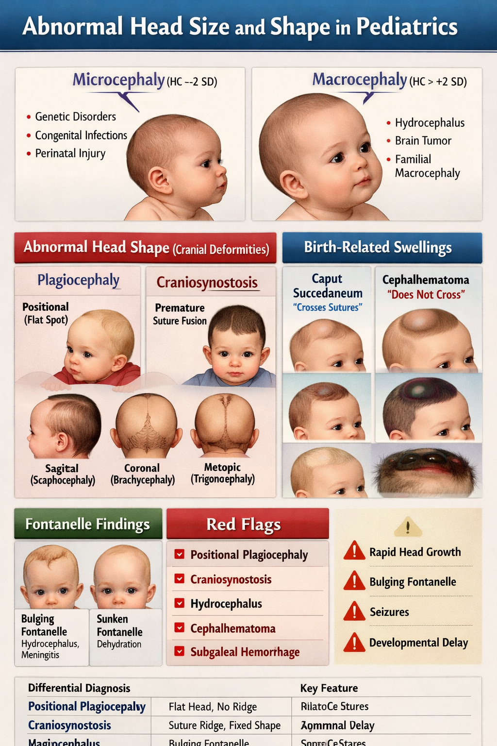
Head circumference is abnormal when it falls below –2 standard deviations (microcephaly) or above +2 standard deviations (macrocephaly) for age and sex on standardized growth charts.

What is microcephaly in children?

Microcephaly is defined as a head circumference less than –2 SD below the mean, indicating reduced brain growth and often associated with developmental delay or neurologic impairment.

What are the common causes of microcephaly?

Common causes include congenital infections (CMV, Zika), chromosomal abnormalities, genetic syndromes, perinatal hypoxic injury, severe malnutrition, and metabolic disorders.

What is macrocephaly in pediatrics?

Macrocephaly is defined as head circumference greater than +2 SD above the mean. It may be benign familial or due to serious intracranial pathology such as hydrocephalus.

What are the major causes of macrocephaly?

Causes include benign familial macrocephaly, benign enlargement of subarachnoid spaces (BESS), hydrocephalus, intracranial tumors, subdural hematoma, and storage disorders like Tay-Sachs.

How can benign macrocephaly be differentiated from hydrocephalus?

Benign macrocephaly usually has normal development and stable growth, while hydrocephalus presents with rapid head enlargement, bulging fontanelle, vomiting, sunset eye sign, and raised intracranial pressure.

What is plagiocephaly in infants?

Plagiocephaly refers to asymmetrical flattening of the skull, most commonly positional due to prolonged supine positioning, and is usually benign.

How is positional plagiocephaly different from craniosynostosis?

Positional plagiocephaly has no suture ridging and improves with repositioning, whereas craniosynostosis involves premature suture fusion, palpable ridging, fixed deformity, and may cause raised intracranial pressure.

What is craniosynostosis?

Craniosynostosis is the premature fusion of one or more cranial sutures, leading to abnormal head shape and potential restriction of brain growth.

What are the main types of craniosynostosis and head shapes?

Sagittal fusion causes scaphocephaly, coronal fusion causes brachycephaly or anterior plagiocephaly, metopic fusion causes trigonocephaly, and lambdoid fusion causes posterior plagiocephaly.

What imaging is best for diagnosing craniosynostosis?

CT scan with 3D reconstruction is the gold standard investigation for confirming craniosynostosis and identifying the fused suture.

What is caput succedaneum?

Caput succedaneum is scalp edema present at birth that crosses suture lines and resolves spontaneously within 1–2 days.

What is cephalhematoma and how is it recognized?

Cephalhematoma is a subperiosteal hemorrhage that does not cross suture lines, appears hours after birth, and may lead to neonatal jaundice.

What is subgaleal hemorrhage and why is it dangerous?

Subgaleal hemorrhage is bleeding into the subaponeurotic space, often after vacuum delivery. It can cause massive blood loss, shock, and is a neonatal emergency.

What are red flag signs requiring urgent referral in abnormal head size or shape?

Red flags include rapidly increasing head circumference, bulging fontanelle, seizures, vomiting, developmental regression, suspected craniosynostosis, signs of raised intracranial pressure, or suspected abusive head trauma.

MCQ Test - Abnormal Head Size and Shape in Pediatrics Microcephaly Macrocephaly Craniosynostosis Guide

Progress:
0/15
Time: 00:00

1 A 2-month-old infant has head circumference < –3 SD, seizures, poor feeding, and CT shows periventricular calcifications. Hearing screening reveals sensorineural deafness. What is the most likely cause of this microcephaly?

Explanation:

Congenital CMV classically causes severe microcephaly, seizures, periventricular calcifications, and sensorineural hearing loss.

2 A newborn presents with severe microcephaly, intracranial calcifications, and maternal history of fever and rash during pregnancy. The infant also has chorioretinitis. Which infection is most consistent?

Explanation:

Congenital toxoplasmosis causes chorioretinitis, hydrocephalus, and diffuse intracranial calcifications with microcephaly.

3 A 6-month-old infant’s head circumference is rapidly increasing, crossing percentiles, with bulging fontanelle and sunset eye sign. What is the most likely diagnosis?

Explanation:

Rapid head growth with bulging fontanelle and sunset sign strongly indicates hydrocephalus with raised ICP.

4 A 9-month-old has progressive macrocephaly, developmental regression, exaggerated startle response, and a cherry-red spot on fundoscopy. What is the most likely diagnosis?

Explanation:

Tay-Sachs is a lysosomal storage disorder causing macrocephaly, neurodegeneration, and cherry-red spot.

5 A 4-month-old has macrocephaly, normal milestones, and family history of large head size. Cranial ultrasound shows enlarged subarachnoid spaces without ventricular dilation. Best diagnosis?

Explanation:

BESS presents with macrocephaly, normal development, familial tendency, and enlarged subarachnoid spaces.

6 A 3-month-old has occipital flattening, anterior displacement of the ear, and no palpable suture ridge. What is the most likely diagnosis?

Explanation:

Positional plagiocephaly shows anterior ear shift and no suture ridging, improving with repositioning.

7 A 5-month-old has posterior plagiocephaly with ear displaced posteriorly and a palpable ridge over the lambdoid suture. Diagnosis?

Explanation:

Posterior ear displacement with suture ridging suggests lambdoid craniosynostosis.

8 A newborn has an elongated narrow skull with premature fusion of the sagittal suture. What is this head shape called?

Explanation:

Sagittal synostosis produces scaphocephaly (boat-shaped long narrow skull).

9 A newborn has triangular forehead, hypotelorism, and fused metopic suture. What is the diagnosis?

Explanation:

Metopic synostosis causes trigonocephaly with triangular forehead and close-set eyes.

10 A 1-year-old with craniosynostosis develops vomiting, papilledema, and developmental delay. These findings suggest which complication?

Explanation:

Premature suture fusion can restrict skull expansion leading to increased intracranial pressure.

11 A neonate has scalp swelling present at birth that crosses suture lines and resolves within 48 hours. Diagnosis?

Explanation:

Caput succedaneum is superficial edema crossing sutures and resolves quickly.

12 A neonate develops swelling hours after birth confined to one parietal bone, not crossing sutures, with risk of jaundice. Diagnosis?

Explanation:

Cephalhematoma is subperiosteal, limited by sutures, and may cause hyperbilirubinemia.

13 A vacuum-assisted delivery newborn develops diffuse boggy scalp swelling crossing sutures, pallor, tachycardia, and shock. Most likely diagnosis?

Explanation:

Subgaleal hemorrhage is a neonatal emergency due to massive blood loss into scalp spaces.

14 A 10-month-old infant has macrocephaly with subdural collections on imaging and no clear trauma history. What must be strongly considered?

Explanation:

Subdural hematomas without adequate explanation are highly concerning for abusive head trauma.

15 A 6-month-old suspected craniosynostosis requires definitive imaging. What is the gold standard test?

Explanation:

CT with 3D reconstruction is the gold standard for identifying fused sutures in craniosynostosis.

Test Results

0%
0/15
0
Correct Answers
0
Wrong Answers
00:00
Time Taken
0
Skipped
View Exam Mode MCQs (1770643141-index-8-.html)

Related Articles

Comments