Cardiomyopathies Clinical Types Diagnosis and Management Guide

Author: Medical Editorial Team – Board-certified physicians with 10+ years in emergency medicine. Learn more.

Illustration of Cardiomyopathies Clinical Types Diagnosis and Management Guide symptoms

Medical Disclaimer: This is educational content only, not medical advice. Consult a licensed healthcare provider for diagnosis/treatment. Information based on sources like WHO/CDC guidelines (last reviewed: 2026-02-13).

About the Author: Dr. Dinesh, MBBS, is a qualified medical doctor with over [2 years – add your experience] of experience in general medicine As the owner and lead content creator of LearnWithTest.pro, Dr. Dinesh ensures all articles are based on evidence-based guidelines from sources like WHO, CDC, and peer-reviewed journals. This content is for educational purposes only and not a substitute for professional medical advice.

Contact Details: For inquiries or collaborations, reach out at:

All content is reviewed for accuracy and updated regularly (last review: January 10, 2026). We prioritize trustworthiness by citing reliable sources and adhering to medical ethics.

Frequently Asked Questions

What are cardiomyopathies?

Cardiomyopathies are a group of diseases of the heart muscle characterized by structural and functional abnormalities of the myocardium that are not explained by coronary artery disease, hypertension, valvular disease, or congenital heart disease.

What are the main types of cardiomyopathies?

The main types include dilated cardiomyopathy, hypertrophic cardiomyopathy, restrictive cardiomyopathy, arrhythmogenic right ventricular cardiomyopathy, stress-induced (Takotsubo) cardiomyopathy, and left ventricular non-compaction.

What is dilated cardiomyopathy?

Dilated cardiomyopathy is characterized by dilation of one or both ventricles with reduced systolic function, leading to heart failure, arrhythmias, and thromboembolic complications.

What causes dilated cardiomyopathy?

Causes include genetic mutations, viral myocarditis, alcohol abuse, chemotherapeutic drugs, peripartum state, metabolic disorders, nutritional deficiencies, and idiopathic causes.

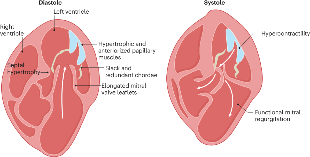
What is hypertrophic cardiomyopathy?

Hypertrophic cardiomyopathy is a genetic disorder characterized by unexplained left ventricular hypertrophy, often involving the interventricular septum, with diastolic dysfunction and possible left ventricular outflow tract obstruction.

Why is hypertrophic cardiomyopathy associated with sudden cardiac death?

Sudden cardiac death occurs due to malignant ventricular arrhythmias, myocardial disarray, ischemia, and dynamic left ventricular outflow tract obstruction, particularly during exertion.

What is restrictive cardiomyopathy?

Restrictive cardiomyopathy is characterized by impaired ventricular filling due to stiff ventricular walls with preserved or near-normal systolic function, leading to predominant diastolic heart failure.

What are common causes of restrictive cardiomyopathy?

Common causes include amyloidosis, sarcoidosis, hemochromatosis, radiation-induced fibrosis, endomyocardial fibrosis, and other infiltrative or storage disorders.

What is arrhythmogenic right ventricular cardiomyopathy?

Arrhythmogenic right ventricular cardiomyopathy is a genetic disorder involving fibrofatty replacement of right ventricular myocardium, leading to ventricular arrhythmias, syncope, and sudden cardiac death.

What are the hallmark ECG findings in ARVC?

Characteristic ECG findings include T-wave inversion in leads V1–V3, epsilon waves, ventricular arrhythmias with left bundle branch block morphology, and prolonged QRS duration in right precordial leads.

What is Takotsubo cardiomyopathy?

Takotsubo cardiomyopathy is a transient stress-induced cardiomyopathy characterized by acute reversible left ventricular systolic dysfunction, often following emotional or physical stress, with normal coronary arteries.

How is left ventricular non-compaction diagnosed?

Left ventricular non-compaction is diagnosed using echocardiography or cardiac MRI showing excessive trabeculations, deep intertrabecular recesses, and a high non-compacted to compacted myocardium ratio.

What investigations are essential in suspected cardiomyopathy?

Essential investigations include ECG, echocardiography, cardiac MRI, Holter monitoring, laboratory tests including BNP, genetic testing in selected cases, and endomyocardial biopsy when indicated.

What are the common complications of cardiomyopathies?

Common complications include heart failure, atrial and ventricular arrhythmias, thromboembolism, stroke, and sudden cardiac death.

How are cardiomyopathies generally managed?

Management includes guideline-directed medical therapy for heart failure, treatment of underlying causes, arrhythmia management, anticoagulation when indicated, device therapy such as ICD or CRT, and heart transplantation in end-stage disease.

When is an implantable cardioverter-defibrillator indicated in cardiomyopathies?

ICD is indicated in patients with reduced ejection fraction (≤35%) despite optimal therapy, survivors of cardiac arrest, sustained ventricular tachyarrhythmias, and high-risk hypertrophic or arrhythmogenic cardiomyopathy patients.

Can cardiomyopathies be reversed?

Some cardiomyopathies such as alcohol-induced, peripartum, tachycardia-induced, and stress-induced cardiomyopathies can partially or completely reverse with appropriate treatment and removal of the underlying cause.

How do cardiomyopathies differ from ischemic heart disease?

Cardiomyopathies primarily involve intrinsic myocardial disease, whereas ischemic heart disease results from coronary artery obstruction leading to myocardial ischemia or infarction.

What is the prognosis of cardiomyopathies?

Prognosis varies widely depending on the type, severity, genetic factors, response to therapy, and risk of arrhythmias, ranging from near-normal life expectancy to progressive heart failure requiring transplantation.

Is genetic screening important in cardiomyopathies?

Yes, genetic screening is important in inherited cardiomyopathies such as hypertrophic and arrhythmogenic cardiomyopathy to identify affected family members and guide surveillance and preventive strategies.

MCQ Test - Cardiomyopathies Clinical Types Diagnosis and Management Guide

Progress:
0/15
Time: 00:00

1 A 46-year-old man presents with progressive dyspnea and fatigue. Echocardiography shows a dilated left ventricle, EF 28%, global hypokinesia, and functional mitral regurgitation. Coronary angiography is normal. What is the most likely diagnosis?

Explanation:

Dilated cardiomyopathy is characterized by ventricular dilation, systolic dysfunction, and absence of significant coronary artery disease.

2 A 24-year-old competitive athlete collapses during a football match and cannot be resuscitated. Autopsy shows asymmetric septal hypertrophy and myocardial fiber disarray. What is the most likely mechanism of death?

Explanation:

Hypertrophic cardiomyopathy commonly causes sudden cardiac death due to ventricular arrhythmias, especially during exertion.

3 A patient with dilated cardiomyopathy remains symptomatic despite optimal medical therapy. EF is 32% and Holter monitoring shows non-sustained ventricular tachycardia. What intervention best reduces mortality?

Explanation:

ICD implantation reduces sudden cardiac death in patients with cardiomyopathy and EF ≤35%.

4 A 62-year-old man presents with ascites, hepatomegaly, pedal edema, and raised JVP. Echocardiography shows preserved EF, normal ventricular size, and marked biatrial enlargement with restrictive filling pattern. What is the most likely diagnosis?

Explanation:

Restrictive cardiomyopathy presents with severe diastolic dysfunction, preserved systolic function, and biatrial enlargement.

5 A patient with thickened ventricular walls on echocardiography has low-voltage QRS complexes on ECG. Which condition best explains this finding?

Explanation:

Low-voltage ECG with increased wall thickness is classic for infiltrative cardiomyopathy such as amyloidosis.

6 A 34-year-old woman develops acute chest pain and breathlessness after severe emotional stress. ECG mimics acute coronary syndrome, but coronary angiography is normal. Echocardiography shows apical ballooning. What is the diagnosis?

Explanation:

Takotsubo cardiomyopathy is stress-induced, reversible LV systolic dysfunction with normal coronaries.

7 A 27-year-old man presents with recurrent syncope. ECG shows T-wave inversion in V1–V3 and epsilon waves. Cardiac MRI reveals fibrofatty infiltration of the right ventricle. What is the most likely diagnosis?

Explanation:

ARVC is characterized by fibrofatty replacement of the right ventricle and ventricular arrhythmias.

8 A patient with hypertrophic cardiomyopathy experiences worsening dyspnea. Which drug is most appropriate as first-line therapy?

Explanation:

Beta-blockers reduce heart rate, improve diastolic filling, and reduce LV outflow tract obstruction in HCM.

9 A patient with hypertrophic cardiomyopathy has syncope and a family history of sudden cardiac death. What is the most effective preventive strategy?

Explanation:

ICD implantation is the most effective strategy for preventing sudden cardiac death in high-risk HCM patients.

10 A patient with restrictive cardiomyopathy develops atrial fibrillation. Why does this cause marked clinical deterioration?

Explanation:

In restrictive cardiomyopathy, atrial contraction is crucial for ventricular filling; its loss leads to severe hemodynamic compromise.

11 A patient with dilated cardiomyopathy presents with an ischemic stroke. Echocardiography shows a left ventricular apical thrombus. What is the most appropriate long-term treatment?

Explanation:

LV thrombus in dilated cardiomyopathy requires long-term anticoagulation to prevent embolic events.

12 A young patient has excessive trabeculations and deep intertrabecular recesses on echocardiography. What cardiomyopathy is suggested?

Explanation:

Left ventricular non-compaction results from failure of normal myocardial compaction during development.

13 A patient with chronic alcohol use develops heart failure with reduced ejection fraction. Which intervention has the greatest impact on disease progression?

Explanation:

Alcohol abstinence can significantly improve or reverse alcoholic dilated cardiomyopathy.

14 A patient with dilated cardiomyopathy has a QRS duration of 160 ms with left bundle branch block morphology. Which therapy improves survival and symptoms?

Explanation:

CRT improves ventricular synchrony and outcomes in patients with DCM and wide QRS due to LBBB.

15 A patient with end-stage cardiomyopathy remains symptomatic despite maximal medical and device therapy. What is the definitive treatment?

Explanation:

Heart transplantation is the definitive treatment for refractory end-stage cardiomyopathy.

Test Results

0%
0/15
0
Correct Answers
0
Wrong Answers
00:00
Time Taken
0
Skipped
View Exam Mode MCQs (1767597654-index-2026-01-05T124629.078.html)

Related Articles

Comments