UPPER LIMB ANATOMY – DETAILED STUDY
The upper limb is specialized for mobility, precision, manipulation, and sensation. It connects the axial skeleton to the hand and is divided into regions, bones, joints, muscles, nerves, vessels, and lymphatics.
1. REGIONS OF THE UPPER LIMB
- Pectoral (Shoulder) Region
- Axilla
- Arm (Brachium) – shoulder to elbow
- Forearm (Antebrachium) – elbow to wrist
- Hand (Manus) – wrist to fingers
2. OSTEOLOGY (BONES)
A. Pectoral Girdle
Provides attachment of upper limb to trunk.
- Clavicle
* S-shaped
* Medial end → sternum
* Lateral end → acromion of scapula
* Transmits force from limb to axial skeleton
- Scapula
* Flat triangular bone
* Key landmarks:
* Spine
* Acromion
* Coracoid process
* Glenoid cavity (articulates with humerus)
B. Arm
- Humerus
* Proximal:
* Head
* Anatomical neck
* Surgical neck (fracture site → axillary nerve)
* Greater & lesser tubercles
* Shaft:
* Deltoid tuberosity
* Radial groove (radial nerve)
* Distal:
* Capitulum (radius)
* Trochlea (ulna)
* Medial & lateral epicondyles
C. Forearm
- Radius (lateral)
* Head → elbow rotation
* Styloid process → wrist
- Ulna (medial)
* Olecranon → elbow
* Trochlear notch
* Styloid process
D. Hand
- Carpal bones (8)
Proximal: Scaphoid, Lunate, Triquetrum, Pisiform
Distal: Trapezium, Trapezoid, Capitate, Hamate
- Metacarpals (5)
- Phalanges (14)
3. JOINTS
Shoulder Joint (Glenohumeral)
- Type: Ball and socket
- Movements: Flexion, extension, abduction, adduction, rotation
- Stability provided mainly by muscles, not ligaments
Elbow Joint
- Type: Hinge
- Components:
* Humeroulnar
* Humeroradial
Radioulnar Joints
- Proximal & distal
- Enable pronation and supination
Wrist Joint
- Radiocarpal
- Movements: Flexion, extension, abduction, adduction
4. MUSCLES
A. Shoulder Muscles
Rotator Cuff (SITS)
- Supraspinatus – initiates abduction
- Infraspinatus – lateral rotation
- Teres minor – lateral rotation
- Subscapularis – medial rotation
B. Arm Muscles
Anterior Compartment (Flexors)
- Biceps brachii
- Brachialis
- Coracobrachialis
Nerve: Musculocutaneous
Posterior Compartment (Extensor)
- Triceps brachii
Nerve: Radial
C. Forearm Muscles
Anterior Compartment
- Superficial flexors
- Deep flexors
Nerve: Median (except FCU & medial FDP → ulnar)
Posterior Compartment
- Extensors and supinators
Nerve: Radial
D. Hand Muscles
- Thenar muscles – thumb movements
- Hypothenar muscles – little finger
- Interossei
* Palmar – adduction
* Dorsal – abduction
- Lumbricals – flex MCP, extend IP joints
5. NERVE SUPPLY
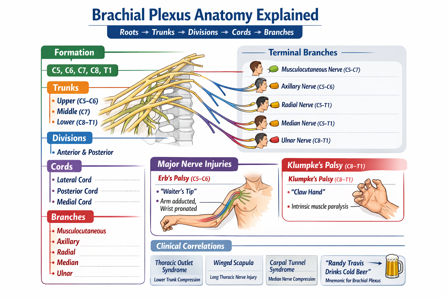
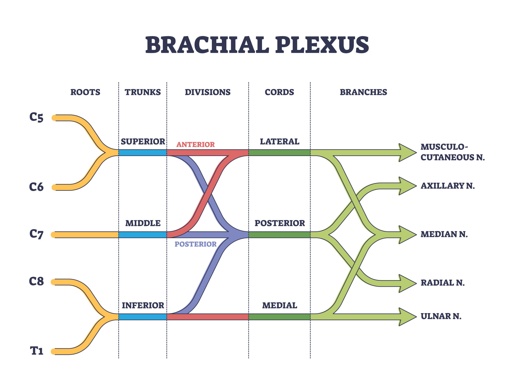
BRACHIAL PLEXUS (C5–T1)
Roots → Trunks → Divisions → Cords → Branches
Major Nerves
- Musculocutaneous – arm flexors
- Axillary – deltoid, teres minor
- Radial – extensors
- Median – most forearm flexors, thenar muscles
- Ulnar – intrinsic hand muscles
6. ARTERIAL SUPPLY
- Subclavian artery
- Axillary artery
- Brachial artery
- Divides into:
* Radial artery
* Ulnar artery
- Palmar arches:
* Superficial
* Deep
7. VENOUS DRAINAGE
Superficial Veins
- Cephalic
- Basilic
- Median cubital (venepuncture)
Deep Veins
- Paired venae comitantes
8. LYMPHATIC DRAINAGE
- Superficial → Axillary nodes
- Deep → Apical nodes → Subclavian trunk
9. FASCIA & SPACES
- Deep fascia forms compartments
- Intermuscular septa
- Clinical relevance:
* Compartment syndrome
* Infections spreading along fascial planes
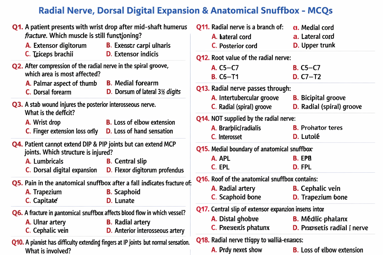
10. CLINICAL CORRELATIONS
- Surgical neck fracture → Axillary nerve injury
- Mid-shaft humerus fracture → Radial nerve palsy
- Carpal tunnel syndrome → Median nerve compression
- Claw hand → Ulnar nerve injury
- Wrist drop → Radial nerve lesion
11. FUNCTIONAL SUMMARY
- Proximal limb → Stability and power
- Distal limb (hand) → Precision and fine movements
- High sensory representation in cortex