Breast Anatomy (Mammary Gland)
1. Definition and Overview
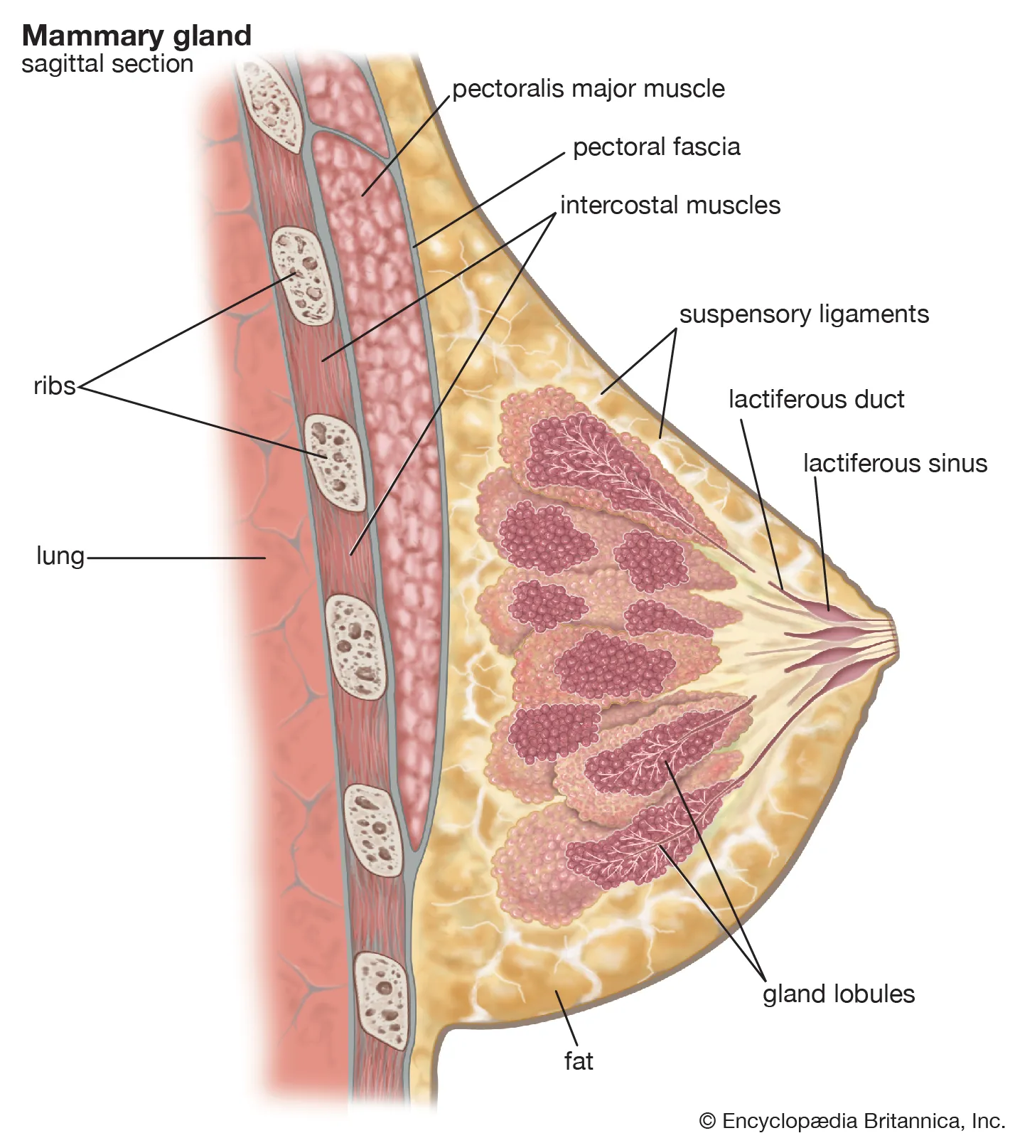
The breast (mammary gland) is a modified apocrine sweat gland, specialized for lactation. It is present in both sexes but is functionally developed in females after puberty under hormonal influence.
2. Location and Extent
- Lies in the superficial fascia of the anterior chest wall
- Vertical extent: 2nd to 6th ribs
- Horizontal extent: Lateral border of sternum → mid-axillary line
- Axillary tail (Tail of Spence): Extension into axilla through foramen of Langer (important in breast cancer spread)
3. External Features
(A) Nipple
- Located at the level of the 4th intercostal space in nulliparous females
- Contains 15–20 openings of lactiferous ducts
- No fat, hair, or sweat glands
- Rich in smooth muscle → erection with stimulation
(B) Areola
- Circular pigmented area around nipple
- Contains Montgomery’s tubercles (modified sebaceous glands)
- Darkens during pregnancy
4. Internal Structure (Gross Anatomy)
(A) Lobes and Lobules
- 15–20 lobes arranged radially
- Each lobe → subdivided into lobules
- Lobules contain alveoli (acini) → milk-secreting units
(B) Duct System
- Alveoli → intralobular ducts → interlobular ducts
- Each lobe drains via a lactiferous duct
- Lactiferous duct shows slight dilatation near nipple (lactiferous sinus – minimal in humans)
5. Supporting Structures
(A) Fibrous Tissue
- Suspensory ligaments of Cooper
- Extend from skin → deep fascia
- Maintain breast shape
- Shortening causes skin dimpling (important sign in carcinoma)
(B) Fat
- Determines size and shape of breast
- Absent beneath nipple and areola
6. Relations
Anterior
- Skin and superficial fascia
Posterior
- Pectoral fascia over:
* Pectoralis major
* Serratus anterior (laterally)
- Retromammary space: Allows mobility of breast over chest wall
7. Blood Supply
Arterial Supply
- Internal thoracic artery (perforating branches)
- Lateral thoracic artery
- Thoracoacromial artery
- Posterior intercostal arteries
Venous Drainage
- Axillary vein
- Internal thoracic vein
8. Lymphatic Drainage (Very Important)
- 75% → Axillary lymph nodes
* Anterior (pectoral)
* Central
* Apical
- Medial quadrants → Parasternal nodes
- Some drainage → posterior intercostal nodes
- Subareolar plexus (Sappey’s plexus) plays key role
9. Nerve Supply
- Intercostal nerves T2–T6
- Nipple mainly supplied by T4
- Sensory supply to skin and nipple
- Sympathetic fibers to blood vessels and smooth muscle
10. Developmental Anatomy
- Develops from mammary ridge (milk line) extending from axilla to groin
- Persistence → accessory breast or nipple (polythelia, polymastia)
11. Changes with Age and Hormones
Puberty
- Estrogen → ductal growth
- Progesterone → lobuloalveolar development
Pregnancy
- Proliferation of alveoli
- Increased vascularity
Lactation
- Prolactin → milk secretion
- Oxytocin → milk ejection
Menopause
- Glandular tissue replaced by fat
- Breast becomes less dense
12. Applied Anatomy (Clinical Importance)
- Breast carcinoma spreads via lymphatics
- Peau d’orange: Lymphatic obstruction
- Retraction of nipple: Involvement of lactiferous ducts
- Mastitis: Infection during lactation
- Gynecomastia: Male breast enlargement
13. Key Exam Points (Quick Recall)
- Tail of Spence → axillary extension
- Cooper’s ligaments → skin dimpling
- Main lymph drainage → axillary nodes
- Nipple nerve supply → T4
- Milk line anomalies → accessory breast/nipple