SCALP AND FACE ANATOMY – COMPLETE DETAILED GUIDE
PART A: SCALP ANATOMY
1. Definition
The scalp is the soft tissue covering the cranial vault, extending:
- Anteriorly: up to the supraorbital margins
- Posteriorly: to the superior nuchal lines
- Laterally: to the zygomatic arches
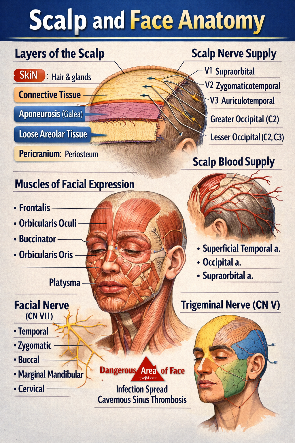
2. Layers of the Scalp (Mnemonic: SCALP)
- S – Skin
* Thick, hair-bearing
* Rich in sebaceous and sweat glands
* Contains hair follicles
* Highly vascular → profuse bleeding from cuts
- C – Connective Tissue (Dense)
* Fibrofatty layer
* Contains blood vessels and nerves
* Vessels are fixed → cannot retract → excessive bleeding
- A – Aponeurosis (Galea Aponeurotica)
* Tough fibrous sheet
* Connects:
* Frontalis muscle (anterior)
* Occipitalis muscle (posterior)
* Lacerations here gape widely
- L – Loose Areolar Tissue
* Also called “danger area of scalp”
* Allows movement of upper 3 layers
* Contains emissary veins → connects scalp veins to intracranial venous sinuses
* Infection may spread → cavernous sinus thrombosis / meningitis
- P – Pericranium
* Periosteum covering skull bones
* Loosely attached except at sutures
* Subperiosteal hematoma limited by sutures
3. Muscles of the Scalp
Occipitofrontalis muscle
- Frontal belly: elevates eyebrows, wrinkles forehead
- Occipital belly: retracts scalp
- Innervation: Facial nerve (CN VII)
4. Blood Supply of Scalp
Arteries (ECA + ICA branches)
- From External Carotid Artery
* Superficial temporal artery
* Posterior auricular artery
* Occipital artery
- From Internal Carotid Artery (Ophthalmic branch)
* Supraorbital artery
* Supratrochlear artery
5. Venous Drainage
- Superficial temporal vein
- Posterior auricular vein
- Occipital vein
→ drain into external jugular vein
Emissary veins
- Connect extracranial veins to intracranial sinuses
- Pathway for infection spread
6. Nerve Supply of Scalp
Sensory (Trigeminal + Cervical nerves)
- Anterior to auricle
* Supraorbital nerve (V1)
* Supratrochlear nerve (V1)
* Zygomaticotemporal nerve (V2)
* Auriculotemporal nerve (V3)
- Posterior to auricle
* Greater occipital nerve (C2)
* Lesser occipital nerve (C2)
* Third occipital nerve (C3)
Motor
- Facial nerve (CN VII) → occipitofrontalis
7. Applied Anatomy of Scalp
- Scalp wounds bleed profusely
- Loose areolar tissue → danger area
- Cephalhematoma (subperiosteal)
- Caput succedaneum (superficial swelling)
PART B: FACE ANATOMY
1. Definition
The face is the anterior part of the head extending:
- From the hairline to the chin
- Between the ears laterally
2. Muscles of Facial Expression
- Derived from second pharyngeal arch
- Insert into skin → facial expressions
- Supplied by Facial nerve (CN VII)
Major Groups
Orbital group
- Orbicularis oculi (closes eye)
Oral group
- Orbicularis oris (closes mouth)
- Buccinator (cheek muscle)
Nasal group
- Nasalis
- Levator labii superioris alaeque nasi
3. Blood Supply of Face
Arteries (External Carotid mainly)
- Facial artery
- Superficial temporal artery
- Maxillary artery
Dangerous area of face
- Upper lip, nose, medial cheek
- Infection may spread via angular vein → cavernous sinus
4. Venous Drainage of Face
- Facial vein
- Angular vein
- Retromandibular vein
Communicates with cavernous sinus via
- Ophthalmic veins
5. Nerve Supply of Face
Sensory – Trigeminal Nerve (CN V)
- Ophthalmic (V1): forehead, upper eyelid
- Maxillary (V2): cheek, upper lip
- Mandibular (V3): lower lip, chin
Motor – Facial Nerve (CN VII)
Branches within parotid gland:
- Temporal
- Zygomatic
- Buccal
- Marginal mandibular
- Cervical
(Mnemonic: To Zanzibar By Motor Car)
6. Lymphatic Drainage of Face
- Submental nodes (chin, lower lip)
- Submandibular nodes (cheeks, upper lip)
- Preauricular nodes (lateral face)
7. Skin of Face
- Thin, highly vascular
- Rich sebaceous glands
- Heals well with minimal scarring
8. Applied Anatomy of Face
- Bell’s palsy → facial nerve paralysis
- Trigeminal neuralgia
- Cavernous sinus thrombosis
- Facial nerve injury during parotid surgery
- Acne common due to sebaceous glands
QUICK EXAM SUMMARY
- Scalp layers: Skin, Connective tissue, Aponeurosis, Loose areolar tissue, Pericranium
- Danger areas: Loose areolar tissue of scalp, Central face
- Motor nerve of face: Facial nerve (CN VII)
- Sensory nerve of face: Trigeminal nerve (CN V)
- Main artery of face: Facial artery