Infratemporal Fossa
Definition
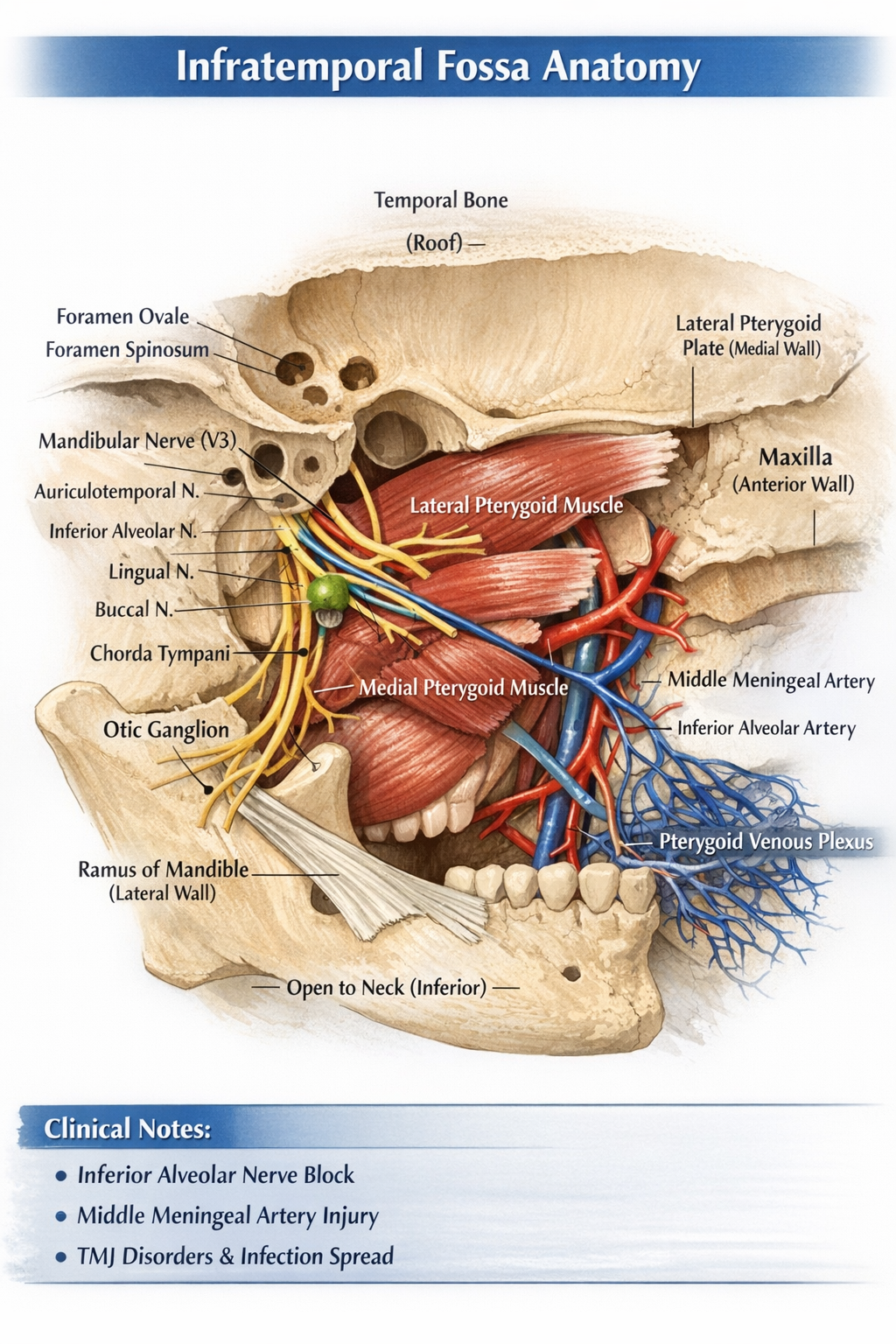
The infratemporal fossa is an irregular, wedge-shaped deep space located below the base of the skull, medial to the ramus of the mandible, and posterior to the maxilla. It is a major neurovascular and muscular compartment involved in mastication, facial sensation, and cranial–facial circulation.
Boundaries
Roof (Superior)
- Infratemporal surface of greater wing of sphenoid
- Squamous part of temporal bone
- Contains:
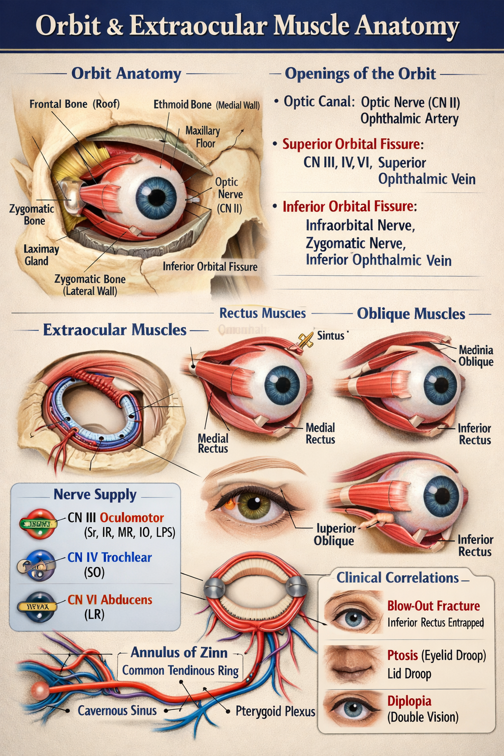
* Foramen ovale – mandibular nerve (V3), accessory meningeal artery, lesser petrosal nerve
* Foramen spinosum – middle meningeal artery and vein, meningeal branch of V3
Lateral Wall
- Ramus of mandible
Medial Wall
- Lateral pterygoid plate (sphenoid)
- Tensor veli palatini muscle
Anterior Wall
- Posterior surface of maxilla
Posterior Wall
- Tympanic plate
- Mastoid and styloid processes of temporal bone
- Separates infratemporal fossa from parotid region
Inferior Boundary
- Open (continuous with tissues of the neck)
Contents
Muscles of Mastication
- Medial pterygoid
* Origin: medial surface of lateral pterygoid plate
* Insertion: medial surface of mandibular angle
* Action: elevation and protrusion of mandible
- Lateral pterygoid
* Two heads:
* Superior: greater wing of sphenoid
* Inferior: lateral pterygoid plate
* Insertion: neck of mandible and TMJ disc
* Action: depression, protrusion, lateral movement of mandible
- Lower part of temporalis (tendon passing through fossa)
Nerves
Mandibular Nerve (V3) – Main Nerve
Exits skull via foramen ovale
Branches
- Auriculotemporal nerve – sensation to temporal region and TMJ
- Inferior alveolar nerve
* Gives nerve to mylohyoid
* Enters mandibular foramen
- Lingual nerve – general sensation to anterior 2/3 of tongue
- Buccal nerve – sensory to cheek
- Motor branches to muscles of mastication
Chorda Tympani (Facial nerve branch)
- Joins lingual nerve
- Carries:
* Taste fibers (anterior 2/3 of tongue)
* Parasympathetic fibers to submandibular and sublingual glands
Otic Ganglion
- Parasympathetic ganglion
- Related to mandibular nerve
- Secretomotor supply to parotid gland via auriculotemporal nerve
Arteries
Maxillary Artery (Terminal branch of external carotid)
Major arterial content of infratemporal fossa
Branches (Mandibular Part)
- Deep auricular artery
- Anterior tympanic artery
- Middle meningeal artery
- Accessory meningeal artery
- Inferior alveolar artery
Veins
Pterygoid Venous Plexus
- Extensive venous network
- Drains into maxillary vein
- Communicates with:
* Cavernous sinus (via emissary veins)
- Clinical relevance: spread of infection
Ligaments
- Sphenomandibular ligament
* Extends from spine of sphenoid to lingula of mandible
* Separates inferior alveolar nerve from lingual nerve
Communications
| Communication | Opening | Connects With |
| --------------------- | ------------------------ | ------------------------------ |
| Middle cranial fossa | Foramen ovale | V3, lesser petrosal nerve |
| Middle cranial fossa | Foramen spinosum | Middle meningeal artery |
| Pterygopalatine fossa | Pterygomaxillary fissure | Maxillary nerve and vessels |
| Orbit | Inferior orbital fissure | Infraorbital nerve and vessels |
| Temporal fossa | Deep to zygomatic arch | Temporalis muscle |
Clinical Importance
- Inferior alveolar nerve block administered in infratemporal fossa
- Middle meningeal artery injury → extradural hematoma
- TMJ disorders involve lateral pterygoid
- Spread of infection via pterygoid venous plexus to cavernous sinus
- Trigeminal neuralgia may involve V3 branches