Radial Nerve, Dorsal Digital Expansion, and Anatomical Snuffbox – Complete Anatomy Guide
1. Radial Nerve
Definition
The radial nerve is the largest branch of the posterior cord of the brachial plexus, providing motor supply to extensor muscles of the upper limb and sensory supply to the posterior arm, forearm, and dorsum of the hand.
Root Value
- C5–T1
Origin
- Arises from the posterior cord of the brachial plexus
Course
Axilla
- Lies posterior to the axillary artery
- Exits the axilla through the lower triangular space
- Accompanied by the profunda brachii artery
Arm
- Enters the radial (spiral) groove of the humerus
- Runs between medial and lateral heads of triceps
- Supplies triceps brachii
- Pierces the lateral intermuscular septum
- Enters the anterior compartment near the lateral epicondyle
Cubital Fossa
- Lies anterior to the lateral epicondyle
- Divides into:
* Superficial branch (sensory)
* Deep branch (motor → posterior interosseous nerve)
Forearm
- Deep branch:
* Pierces supinator
* Continues as posterior interosseous nerve
* Supplies extensor muscles
- Superficial branch:
* Runs under brachioradialis
* Becomes superficial near wrist
* Supplies dorsum of hand
Branches
Motor
- Triceps brachii
- Anconeus
- Brachioradialis
- Extensor carpi radialis longus
- All extensors of wrist and fingers (via posterior interosseous nerve)
Sensory
- Posterior cutaneous nerve of arm
- Posterior cutaneous nerve of forearm
- Dorsal digital nerves to:
* Lateral 3½ digits (proximal parts)
Applied Anatomy
- Radial nerve palsy:
* Wrist drop
* Loss of finger extension
- Mid-shaft humerus fracture → injury in radial groove
- Saturday night palsy → compression in spiral groove
2. Dorsal Digital Expansion (Extensor Expansion / Hood)
Definition
The dorsal digital expansion is a triangular aponeurotic expansion on the dorsum of fingers, formed mainly by extensor digitorum tendon, allowing coordinated finger movements.
Formation
Formed by:
- Extensor digitorum tendon
- Contributions from:
* Lumbricals
* Interossei
Structure
- Central slip:
* Inserts into base of middle phalanx
- Two lateral bands:
* Reunite to insert into distal phalanx
- Covers the dorsum of MCP, PIP, and DIP joints
Functions
- Extension at:
* Metacarpophalangeal (MCP) joints
* Interphalangeal (IP) joints
- Allows interossei and lumbricals to:
* Flex MCP
* Extend IP joints
Clinical Importance
- Mallet finger: rupture of terminal tendon
- Boutonnière deformity: rupture of central slip
- Claw hand deformity: imbalance of intrinsic muscles
3. Anatomical Snuffbox
Definition
The anatomical snuffbox is a triangular depression seen on the lateral aspect of the dorsum of the hand when the thumb is extended.
Boundaries
Lateral (Anterior) Boundary
- Abductor pollicis longus
- Extensor pollicis brevis
Medial (Posterior) Boundary
- Extensor pollicis longus
Proximal Boundary
- Styloid process of radius
Floor
- Scaphoid
- Trapezium
Roof
- Skin
- Superficial fascia
- Superficial branch of radial nerve
- Cephalic vein
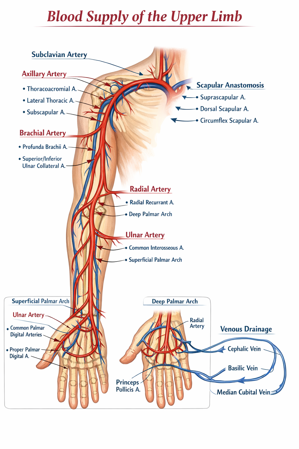
Contents
- Radial artery (main content)
Clinical Importance
- Scaphoid fracture:
* Tenderness in snuffbox
* Risk of avascular necrosis
- Radial artery palpation site
- IV cannulation landmark
Quick Exam Correlation Table
| Structure | Key Clinical Point |
| ------------------------ | ----------------------------- |
| Radial nerve | Wrist drop |
| Dorsal digital expansion | Finger extension coordination |
| Anatomical snuffbox | Scaphoid fracture tenderness |