Orbit and Extraocular Muscle Anatomy
1. Orbit – Overview
The orbit is a paired pyramidal bony cavity that houses and protects the eyeball and its associated structures.
Contents
- Eyeball (globe)
- Extraocular muscles
- Optic nerve (CN II)
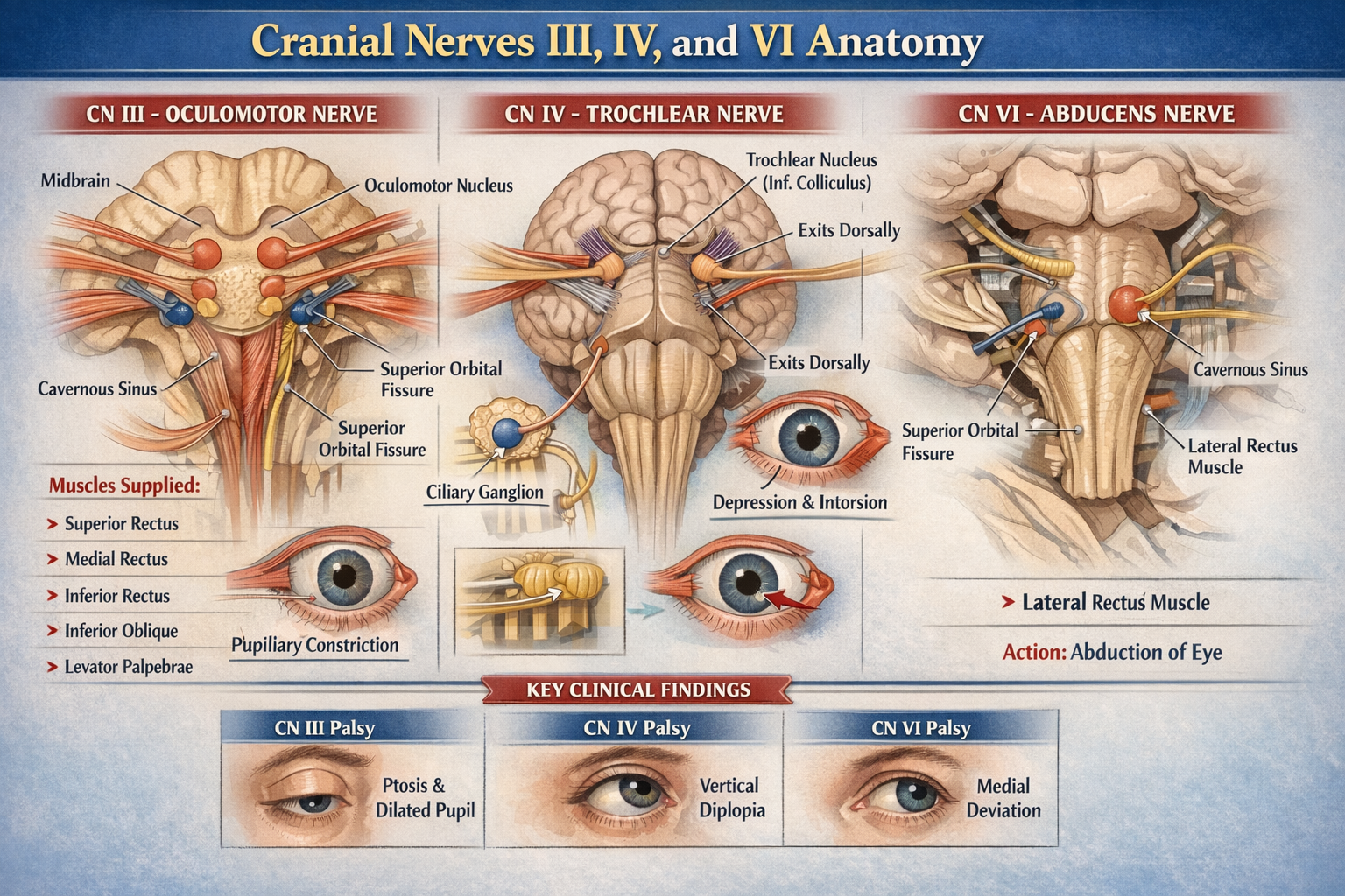
- Oculomotor (CN III), Trochlear (CN IV), Abducens (CN VI)
- Ophthalmic division of trigeminal nerve (CN V1)
- Lacrimal gland
- Ophthalmic artery and veins
- Orbital fat and connective tissue
Shape and Orientation
- Pyramidal: base anterior (orbital margin), apex posterior (optic canal)
- Apex points medially and posteriorly
2. Bony Walls of the Orbit
Roof
Bones: Frontal bone, lesser wing of sphenoid
Relations: Anterior cranial fossa, frontal sinus
Structures: Lacrimal gland fossa, trochlear fossa
Floor
Bones: Maxilla, zygomatic, palatine
Relations: Maxillary sinus
Clinical: Most common site of blow-out fracture
Structures: Infraorbital groove and canal
Medial Wall
Bones: Ethmoid (lamina papyracea), lacrimal, maxilla, sphenoid
Relations: Ethmoidal air sinuses
Clinical: Very thin → spread of infection
Lateral Wall
Bones: Zygomatic, greater wing of sphenoid
Strongest wall
Relations: Temporal fossa
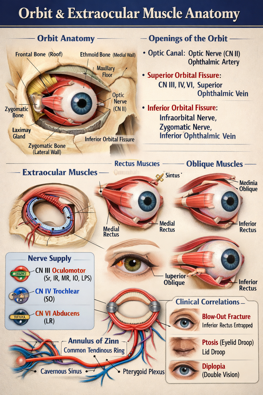
3. Openings of the Orbit
Optic Canal
- Contents: Optic nerve (CN II), ophthalmic artery
- Location: Lesser wing of sphenoid
Superior Orbital Fissure
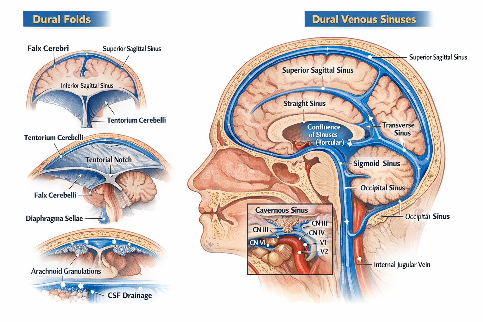
- Contents: CN III, IV, V1, VI, superior ophthalmic vein
- Connects: Middle cranial fossa
Inferior Orbital Fissure
- Contents: Infraorbital nerve, zygomatic nerve, inferior ophthalmic vein
- Connects: Pterygopalatine and infratemporal fossae
4. Extraocular Muscles – Overview
There are 7 extraocular muscles:
- 4 Recti: Superior, Inferior, Medial, Lateral
- 2 Obliques: Superior, Inferior
- 1 Elevator: Levator palpebrae superioris
Common Origin
- Annulus of Zinn (Common tendinous ring) at orbital apex
(All recti + levator palpebrae superioris)
5. Rectus Muscles
Superior Rectus
- Origin: Annulus of Zinn
- Insertion: Superior sclera (anterior to equator)
- Action: Elevation, adduction, intorsion
- Nerve: Oculomotor nerve (CN III – superior division)
Inferior Rectus
- Origin: Annulus of Zinn
- Insertion: Inferior sclera
- Action: Depression, adduction, extorsion
- Nerve: Oculomotor nerve (CN III – inferior division)
Medial Rectus
- Origin: Annulus of Zinn
- Insertion: Medial sclera
- Action: Adduction
- Nerve: Oculomotor nerve (CN III)
Lateral Rectus
- Origin: Annulus of Zinn
- Insertion: Lateral sclera
- Action: Abduction
- Nerve: Abducens nerve (CN VI)
6. Oblique Muscles
Superior Oblique
- Origin: Body of sphenoid
- Course: Passes through trochlea (fibrous pulley)
- Insertion: Posterosuperolateral sclera
- Action: Intorsion, depression, abduction
- Nerve: Trochlear nerve (CN IV)
Inferior Oblique
- Origin: Anterior orbital floor (maxilla)
- Insertion: Posteroinferolateral sclera
- Action: Extorsion, elevation, abduction
- Nerve: Oculomotor nerve (CN III)
7. Levator Palpebrae Superioris
- Origin: Lesser wing of sphenoid
- Insertion: Upper eyelid
- Action: Elevation of upper eyelid
- Nerve: Oculomotor nerve (CN III – superior division)
- Sympathetic supply: Müller’s muscle (smooth muscle component)
8. Blood Supply
- Ophthalmic artery (branch of internal carotid)
- Supplies eyeball, muscles, optic nerve
Venous Drainage
- Superior and inferior ophthalmic veins
- Drain into cavernous sinus and pterygoid plexus
9. Nerve Supply Summary (Rule of 3-4-6)
- CN III (Oculomotor): All extraocular muscles except SO and LR
- CN IV (Trochlear): Superior oblique
- CN VI (Abducens): Lateral rectus