Neurovascular Structures of the Neck
The neck contains vital arterial, venous, and neural structures that connect the brain with the rest of the body. These structures are arranged in distinct fascial compartments, most importantly within the carotid sheath.
1. Arterial Structures of the Neck
A. Common Carotid Arteries (CCA)
- Origin
* Right: From brachiocephalic trunk
* Left: From arch of aorta
- Course
* Ascend in the neck within the carotid sheath
* Divide at C4 vertebral level (upper border of thyroid cartilage)
- Termination
* External carotid artery
* Internal carotid artery
B. External Carotid Artery (ECA)
- Supplies structures of face, scalp, neck
- Lies anteromedial to internal carotid initially
Branches (Mnemonic: Some Angry Lady Figured Out PMS)
- Superior thyroid
- Ascending pharyngeal
- Lingual
- Facial
- Occipital
- Posterior auricular
- Maxillary
- Superficial temporal
C. Internal Carotid Artery (ICA)
- Supplies brain and eye
- No branches in the neck
- Enters skull via carotid canal
- Clinical importance: stroke, carotid stenosis
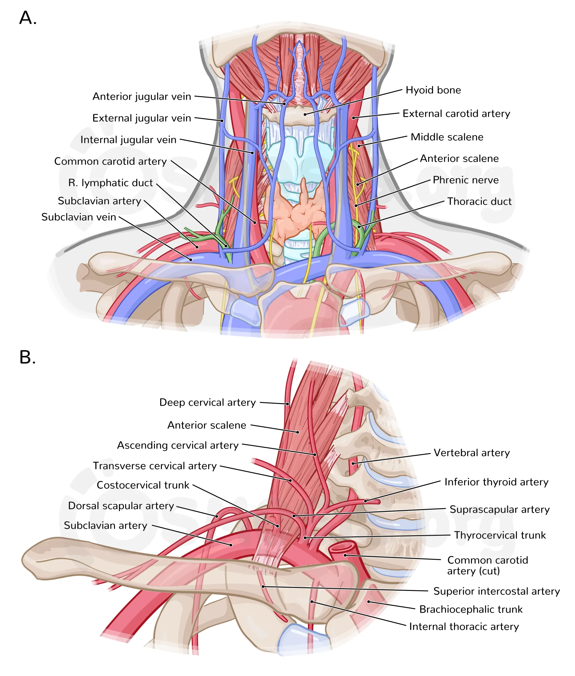
D. Subclavian Artery (Neck Part)
Major branches
- Vertebral artery → brainstem, posterior brain
- Thyrocervical trunk
- Costocervical trunk
2. Venous Structures of the Neck
A. Internal Jugular Vein (IJV)
- Drains brain, face, neck
- Lies lateral to common carotid artery
- Joins subclavian vein → brachiocephalic vein
- Important landmark for central venous access
B. External Jugular Vein (EJV)
- Drains scalp and face
- Superficial, crosses sternocleidomastoid
- Visible in raised venous pressure
C. Anterior Jugular Veins
- Drain submental region
- May form jugular venous arch
3. Neural Structures of the Neck
A. Cranial Nerves in the Neck
1. Vagus Nerve (CN X)
- Lies between carotid artery and jugular vein
- Supplies:
* Parasympathetic to thoracic and abdominal organs
* Laryngeal branches (voice)
2. Glossopharyngeal Nerve (CN IX)
- Supplies:
* Stylopharyngeus
* Taste posterior 1/3 tongue
* Carotid body & sinus
3. Accessory Nerve (CN XI)
- Supplies:
* Sternocleidomastoid
* Trapezius
- Vulnerable during neck surgeries
4. Hypoglossal Nerve (CN XII)
- Motor to tongue muscles
- Injury causes tongue deviation
B. Cervical Plexus (C1–C4)
- Located deep to sternocleidomastoid
- Sensory branches
* Lesser occipital
* Great auricular
* Transverse cervical
* Supraclavicular
- Motor branches
* Ansa cervicalis → infrahyoid muscles
* Phrenic nerve (C3–C5) → diaphragm
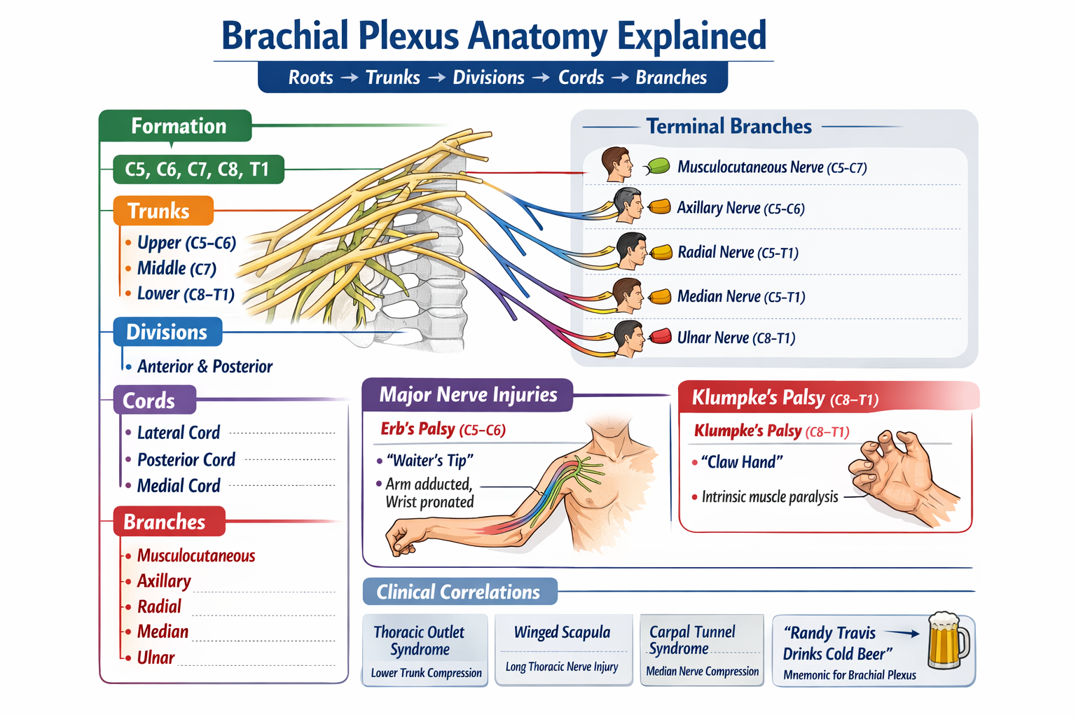
C. Brachial Plexus (Neck Part)
- Roots: C5–T1
- Lies between anterior and middle scalene muscles
- Supplies upper limb
D. Sympathetic Trunk
- Lies posterior to carotid sheath
- Cervical ganglia:
* Superior
* Middle
* Inferior (stellate)
- Injury → Horner syndrome
* Ptosis
* Miosis
* Anhidrosis
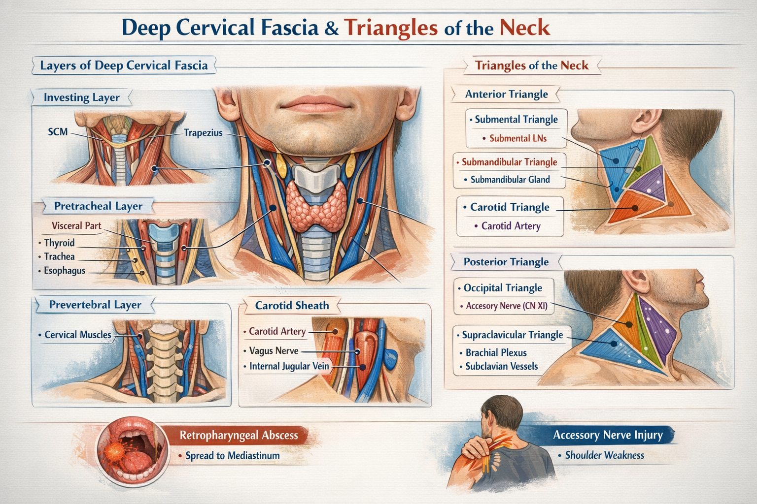
4. Carotid Sheath – Core Neurovascular Bundle
Contents
- Common/Internal carotid artery (medial)
- Internal jugular vein (lateral)
- Vagus nerve (posterior)
- Deep cervical lymph nodes
- Carotid plexus (sympathetic fibers)
Clinical relevance
- Carotid endarterectomy
- Central line placement
- Neck trauma
5. Important Clinical Correlations
- Carotid sinus: Baroreceptor → BP regulation
- Carotid body: Chemoreceptor → oxygen sensing
- Neck hematoma: Can compress airway
- Surgical risk: Accessory nerve injury → shoulder droop
Quick Summary Table
| Structure | Function |
| ----------------- | ------------------------- |
| Carotid arteries | Brain & face blood supply |
| Jugular veins | Venous drainage of head |
| Vagus nerve | Parasympathetic, voice |
| Cervical plexus | Neck sensation & movement |
| Sympathetic trunk | Autonomic control |