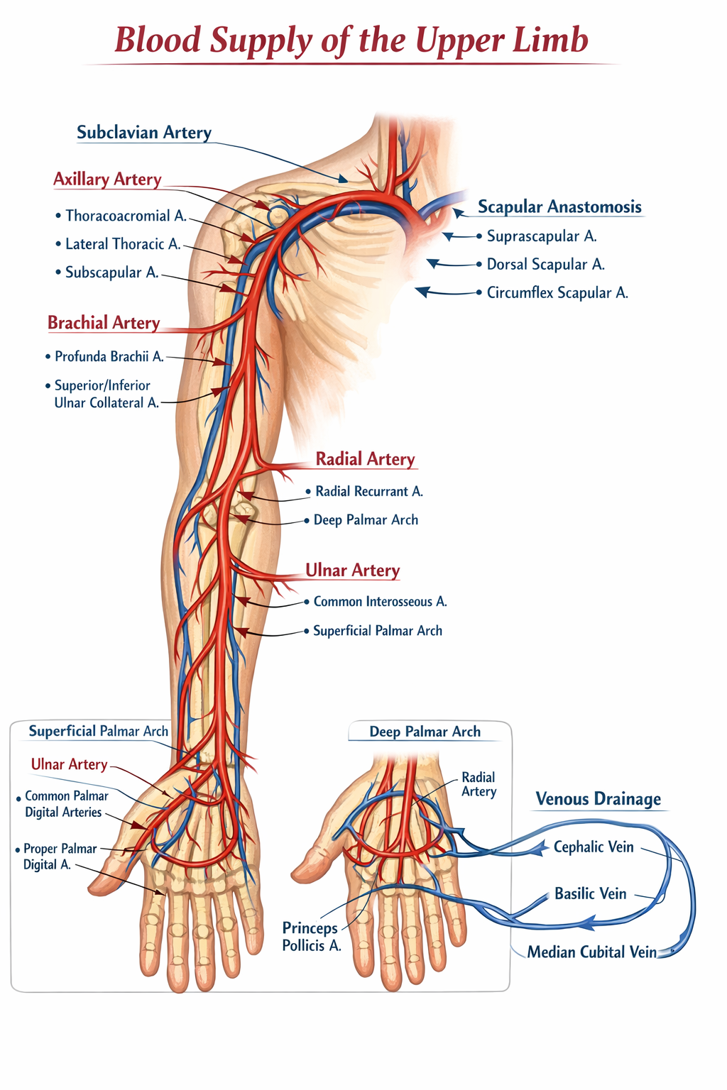
Blood Supply of Upper Limb (Anatomy) – Detailed Explanation
The blood supply of the upper limb is provided mainly by branches of the subclavian artery, which continues as the axillary artery and then the brachial artery, finally dividing into radial and ulnar arteries to supply the forearm and hand.
1. Arterial Supply of Upper Limb
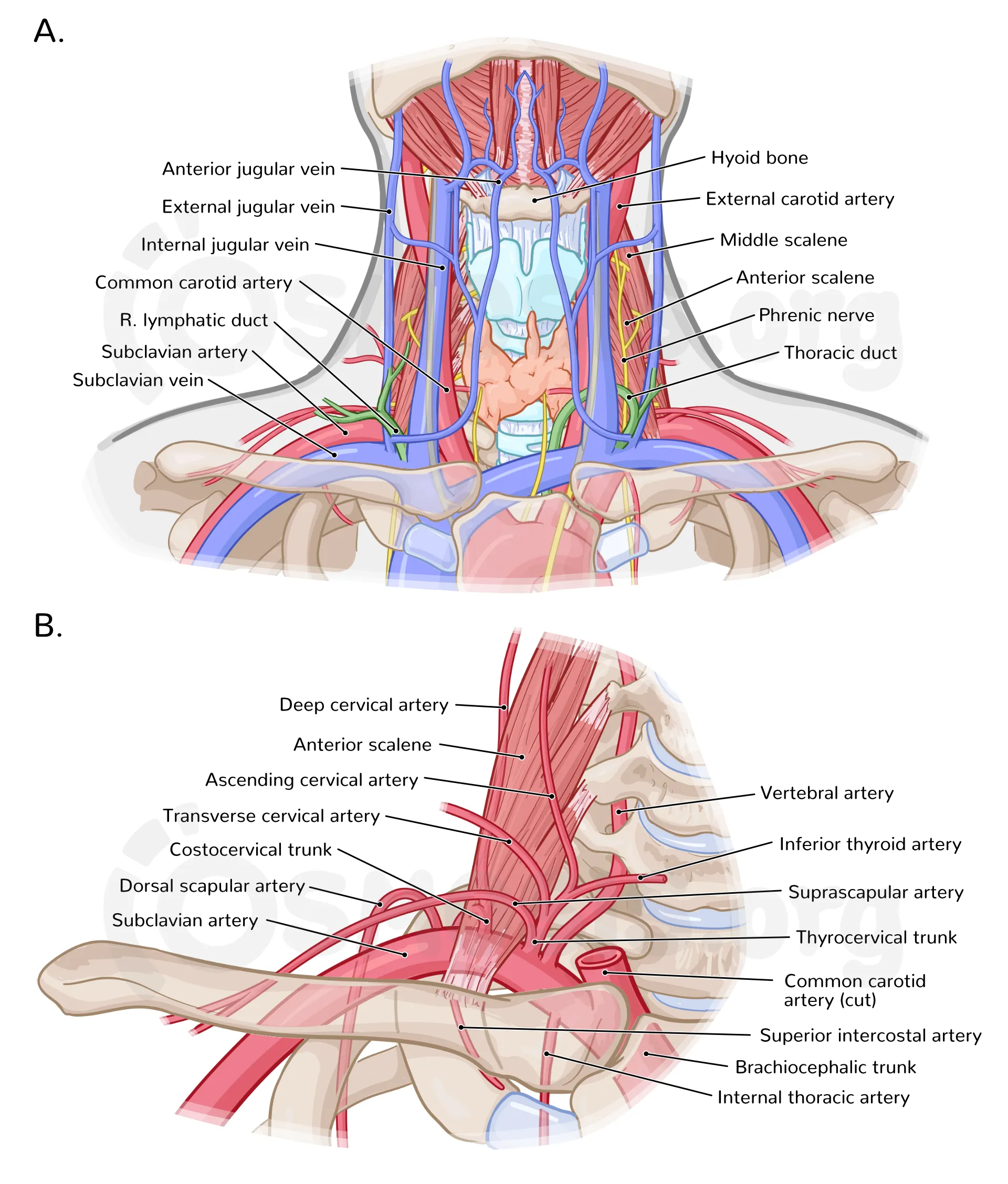
A. Subclavian Artery (Proximal Source)
- Origin:
* Right side: From brachiocephalic trunk
* Left side: From aortic arch
- Supplies the upper limb after crossing the lateral border of the first rib
- Continues as the axillary artery
Important branches related to upper limb
- Thyrocervical trunk (indirect contribution)
- Suprascapular artery (important for scapular anastomosis)
- Dorsal scapular artery (if present)
B. Axillary Artery
- Continuation of subclavian artery at lateral border of 1st rib
- Ends at the lower border of teres major muscle
- Divided into three parts by the pectoralis minor muscle
1st Part (proximal to pectoralis minor)
- Superior thoracic artery
* Supplies upper thoracic wall and first two intercostal spaces
2nd Part (posterior to pectoralis minor)
- Thoracoacromial artery
* Branches: clavicular, acromial, deltoid, pectoral
- Lateral thoracic artery
* Supplies lateral chest wall and breast
3rd Part (distal to pectoralis minor)
- Subscapular artery
* Circumflex scapular artery
* Thoracodorsal artery
- Anterior circumflex humeral artery
- Posterior circumflex humeral artery
* Supplies deltoid and shoulder joint (passes through quadrangular space)
C. Brachial Artery
- Continuation of axillary artery at lower border of teres major
- Runs along medial side of arm
- Ends in cubital fossa by dividing into radial and ulnar arteries
Branches
- Profunda brachii artery
* Travels with radial nerve in spiral groove
* Supplies posterior compartment of arm
- Nutrient artery to humerus
- Superior and inferior ulnar collateral arteries (elbow anastomosis)
- Muscular branches
D. Arteries of Forearm
1. Radial Artery
- Lateral terminal branch of brachial artery
- Runs along lateral forearm
- Palpable at wrist (radial pulse)
- Enters hand through anatomical snuffbox
Branches
- Radial recurrent artery (elbow)
- Muscular branches
- Superficial palmar branch
- Princeps pollicis artery (thumb)
- Radialis indicis artery (index finger)
2. Ulnar Artery
- Medial terminal branch of brachial artery
- Major contributor to palmar arches
Branches
- Anterior and posterior ulnar recurrent arteries
- Common interosseous artery
* Anterior interosseous artery
* Posterior interosseous artery
- Muscular branches
E. Arteries of Hand
Superficial Palmar Arch
- Mainly formed by ulnar artery
- Completed by superficial branch of radial artery
- Gives:
* Common palmar digital arteries
* Proper palmar digital arteries
Deep Palmar Arch
- Mainly formed by radial artery
- Completed by deep branch of ulnar artery
- Gives:
* Palmar metacarpal arteries
* Perforating branches
2. Venous Drainage of Upper Limb
A. Superficial Veins
- Cephalic vein
* Lateral side
* Drains into axillary vein
- Basilic vein
* Medial side
* Joins brachial veins
- Median cubital vein
* Connects cephalic and basilic veins
* Common site for venepuncture
B. Deep Veins
- Paired veins accompanying arteries (venae comitantes)
- Radial veins
- Ulnar veins
- Brachial veins
- Axillary vein → subclavian vein → brachiocephalic vein
3. Important Arterial Anastomoses
Scapular Anastomosis
- Allows collateral circulation during axillary artery blockage
- Involves:
* Suprascapular artery
* Dorsal scapular artery
* Circumflex scapular artery
Elbow Anastomosis
- Between brachial artery branches and forearm recurrent arteries
4. Clinical Importance
- Radial artery used for pulse and arterial blood gas sampling
- Median cubital vein used for venepuncture
- Profunda brachii injury associated with humeral shaft fractures
- Axillary artery injury may compromise upper limb circulation