Cranial Nerves III, IV, and VI – Detailed Anatomy
Overview (Common Function)
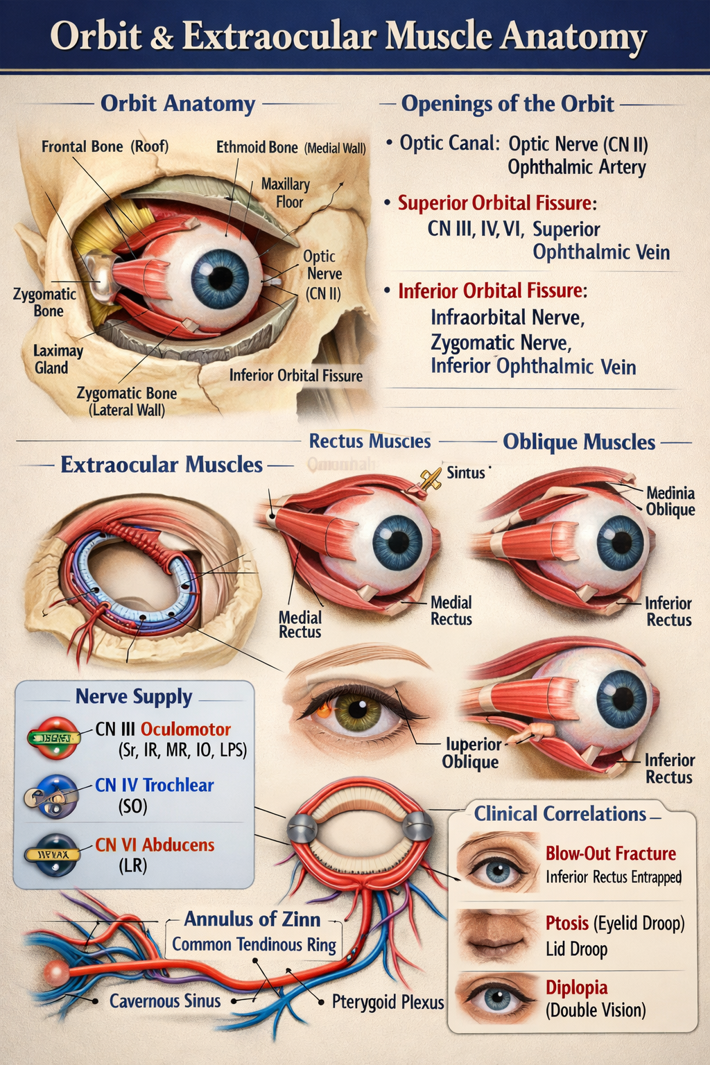
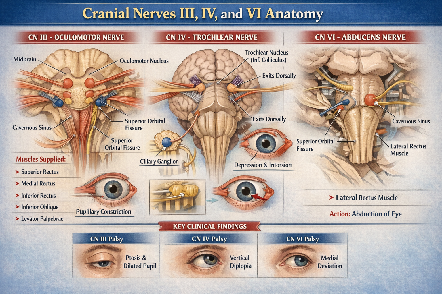
Cranial nerves III, IV, and VI are purely motor nerves supplying the extra-ocular muscles that control eye movements.
Cranial nerve III also carries parasympathetic fibers for pupil constriction and accommodation.
CRANIAL NERVE III – OCULOMOTOR NERVE
Functional Components
- Somatic efferent → extra-ocular muscles
- General visceral efferent (parasympathetic) → pupil and lens
Nuclei (Midbrain)
Located at the level of the superior colliculus:
- Oculomotor nuclear complex
* Supplies all extra-ocular muscles except SO and LR
- Edinger–Westphal nucleus
* Parasympathetic nucleus
* Supplies sphincter pupillae and ciliary muscle
Intracranial Course
- Emerges from ventral midbrain in the interpeduncular fossa
- Passes between:
* Posterior cerebral artery
* Superior cerebellar artery
- Runs in lateral wall of cavernous sinus
- Divides into superior and inferior divisions
- Enters orbit through superior orbital fissure (inside common tendinous ring)
Orbital Course and Branches
Superior Division
- Levator palpebrae superioris
- Superior rectus
Inferior Division
- Medial rectus
- Inferior rectus
- Inferior oblique
- Parasympathetic root to ciliary ganglion
Parasympathetic Pathway
- Edinger–Westphal nucleus → oculomotor nerve
- Synapse in ciliary ganglion
- Postganglionic fibers via short ciliary nerves to:
* Sphincter pupillae → pupillary constriction
* Ciliary muscle → accommodation
Muscles Supplied
- Superior rectus
- Inferior rectus
- Medial rectus
- Inferior oblique
- Levator palpebrae superioris
Actions
- Eye elevation, depression, adduction
- Pupillary constriction
- Accommodation
- Eyelid elevation
CRANIAL NERVE IV – TROCHLEAR NERVE
Functional Component
- Somatic efferent only
Nucleus (Midbrain)
- Located at level of inferior colliculus
Unique Anatomical Features
- Only cranial nerve:
* That emerges dorsally
* That decussates completely
* That has the longest intracranial course
* That supplies the contralateral muscle
Intracranial Course
- Exits dorsal midbrain
- Winds around brainstem laterally
- Passes through:
* Cavernous sinus (lateral wall)
* Superior orbital fissure (outside common tendinous ring)
Orbital Course
- Enters orbit superiorly
- Supplies superior oblique muscle
Muscle Supplied
- Superior oblique
Action of Superior Oblique
- Intorsion
- Depression (especially in adducted eye)
- Abduction
CRANIAL NERVE VI – ABDUCENS NERVE
Functional Component
- Somatic efferent only
Nucleus (Pons)
- Located in dorsal pons, beneath facial colliculus
- Facial nerve fibers loop around abducens nucleus
Intracranial Course
- Emerges at pontomedullary junction
- Ascends along clivus
- Sharp bend over petrous apex
- Passes through cavernous sinus (adjacent to internal carotid artery)
- Enters orbit via superior orbital fissure (inside common tendinous ring)
Orbital Course
- Supplies lateral rectus muscle
Muscle Supplied
- Lateral rectus
Action
- Abduction of eyeball
Summary Table
| Cranial Nerve | Nucleus Level | Exit from Brainstem | Muscle Supplied | Main Action |
| ------------- | -------------------------- | ----------------------- | -------------------------- | -------------------------------- |
| CN III | Midbrain (sup. colliculus) | Ventral | Most EOM + parasympathetic | Eye movement, pupil constriction |
| CN IV | Midbrain (inf. colliculus) | Dorsal | Superior oblique | Depression, intorsion |
| CN VI | Pons | Pontomedullary junction | Lateral rectus | Abduction |
Key Clinical Correlations (Brief)
- CN III palsy → ptosis, dilated pupil, eye down and out
- CN IV palsy → vertical diplopia, worse on stairs
- CN VI palsy → inability to abduct eye, medial deviation