Pharyngeal Arches, Clefts and Pouches
Introduction
Pharyngeal (branchial) arches, clefts, and pouches are transient embryological structures that develop during the 4th–5th week of intrauterine life. They contribute to the formation of the face, neck, pharynx, larynx, and associated neurovascular structures. A clear understanding of their derivatives is essential for anatomy, embryology, ENT, dentistry, and clinical medicine.
Pharyngeal Arches (Branchial Arches)
There are six paired pharyngeal arches (the 5th arch is rudimentary and disappears). Each arch consists of:
- Cartilage (skeletal element)
- Muscle
- Artery (aortic arch derivative)
- Cranial nerve
First Pharyngeal Arch (Mandibular Arch)
Cartilage
- Meckel’s cartilage → malleus, incus
- Mandible (membranous ossification)
Muscles
- Muscles of mastication
- Mylohyoid
- Anterior belly of digastric
- Tensor tympani
- Tensor veli palatini
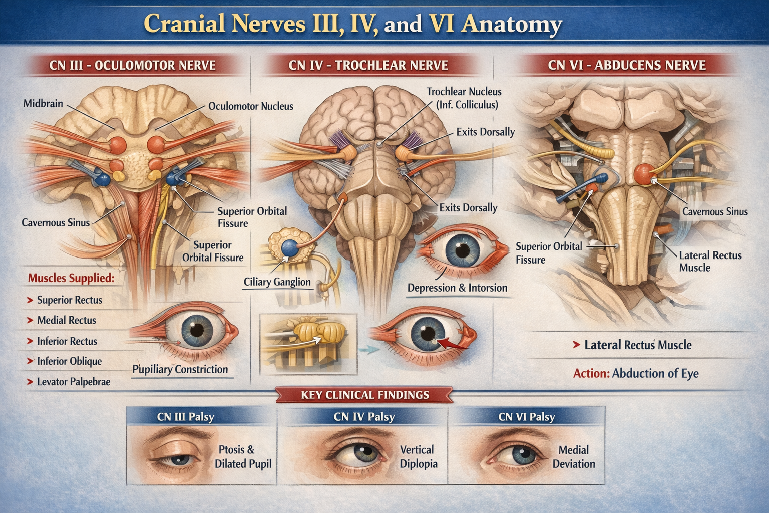
Nerve
- Trigeminal nerve (CN V2, V3)
Artery
- Maxillary artery
Clinical relevance
- Treacher Collins syndrome
- Pierre Robin sequence
Second Pharyngeal Arch (Hyoid Arch)
Cartilage
- Reichert cartilage → stapes, styloid process
- Stylohyoid ligament
- Lesser horn and upper body of hyoid
Muscles
- Muscles of facial expression
- Stapedius
- Stylohyoid
- Posterior belly of digastric
Nerve
- Facial nerve (CN VII)
Artery
- Stapedial artery (regresses)
Clinical relevance
- Facial nerve palsy
- Congenital stapes fixation
Third Pharyngeal Arch
Cartilage
- Greater horn and lower body of hyoid
Muscles
- Stylopharyngeus
Nerve
- Glossopharyngeal nerve (CN IX)
Artery
- Common carotid artery
- Proximal internal carotid artery
Clinical relevance
- Dysphagia due to stylopharyngeus dysfunction
Fourth Pharyngeal Arch
Cartilage
- Laryngeal cartilages (thyroid, cricoid, arytenoid – partially)
Muscles
- Pharyngeal constrictors
- Cricothyroid
- Levator veli palatini
Nerve
- Vagus nerve (CN X) – superior laryngeal nerve
Artery
- Left: part of arch of aorta
- Right: proximal right subclavian artery
Sixth Pharyngeal Arch
Cartilage
- Intrinsic laryngeal cartilages
Muscles
- Intrinsic muscles of larynx (except cricothyroid)
Nerve
- Vagus nerve (CN X) – recurrent laryngeal nerve
Artery
- Pulmonary arteries
- Ductus arteriosus (left)
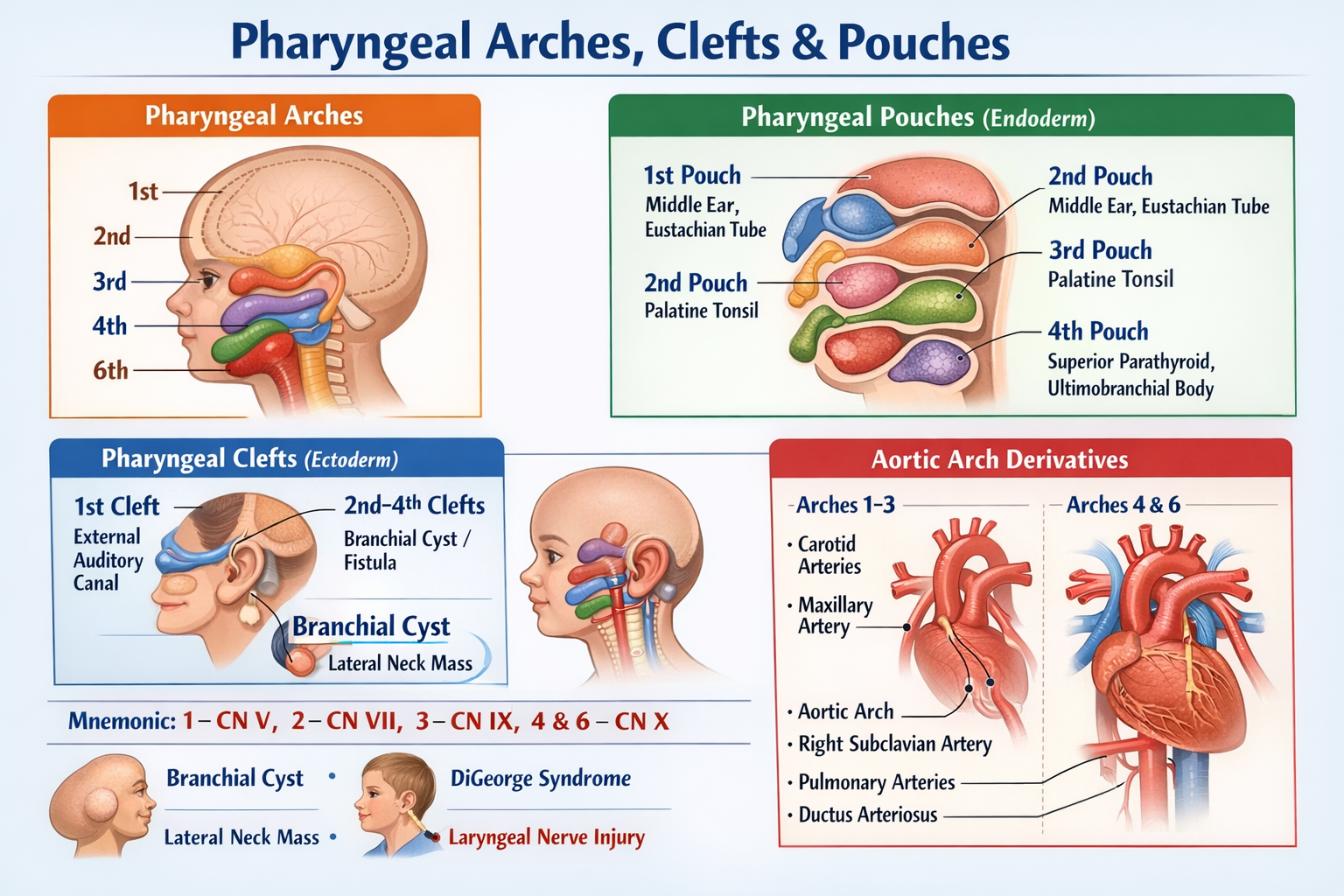
Pharyngeal Clefts (Grooves)
Pharyngeal clefts are ectodermal invaginations between arches.
First Pharyngeal Cleft
- Forms external auditory canal
- Contributes to tympanic membrane
Second, Third and Fourth Clefts
- Normally obliterated
- Persistence leads to branchial cysts, sinuses, or fistulae
Clinical relevance
- Branchial cleft cyst presents as painless lateral neck swelling
Pharyngeal Pouches
Pharyngeal pouches are endodermal outpocketings between arches.
First Pharyngeal Pouch
- Middle ear cavity
- Auditory (Eustachian) tube
Second Pharyngeal Pouch
- Palatine tonsil
- Tonsillar fossa
Third Pharyngeal Pouch
- Dorsal wing → Inferior parathyroid glands
- Ventral wing → Thymus
Fourth Pharyngeal Pouch
- Dorsal wing → Superior parathyroid glands
- Ventral wing → Ultimobranchial body (parafollicular C cells of thyroid)
Clinical relevance
- DiGeorge syndrome (3rd and 4th pouch failure)
Common Exam Memory Aid
Arches → Nerves
- 1 → CN V
- 2 → CN VII
- 3 → CN IX
- 4 & 6 → CN X
Clinical Correlations
- Branchial cyst: Persistent clefts
- DiGeorge syndrome: Thymic aplasia, hypocalcemia
- Recurrent laryngeal nerve injury: Hoarseness of voice
- Congenital ear anomalies: First arch defects
Summary
Pharyngeal arches, clefts, and pouches form the structural and functional framework of the head and neck. Each arch has a distinct nerve, muscle, cartilage, and arterial derivative, while clefts and pouches give rise to ear, tonsils, thymus, parathyroids, and thyroid components. Their abnormalities explain many congenital ENT and neck disorders.