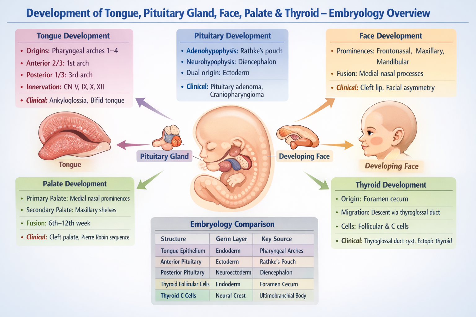
Development of Tongue – Embryology Guide
Embryonic Origin
- Develops from the floor of the primitive pharynx
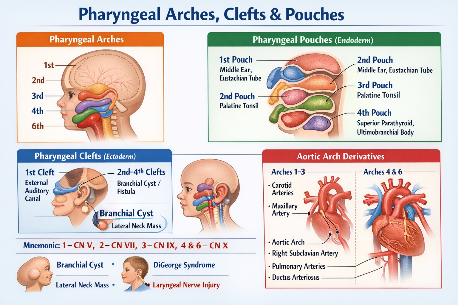
- Derived mainly from pharyngeal arches 1, 2, 3, and 4
- Development begins in 4th week of gestation
Developmental Parts
Anterior two-thirds
- From 1st pharyngeal arch
- Structures:
* Two lateral lingual swellings
* One tuberculum impar
- Lateral swellings overgrow tuberculum impar
- General sensation: Lingual nerve (V3)
Posterior one-third
- From 3rd pharyngeal arch
- Formed by hypobranchial eminence
- Taste + sensation: Glossopharyngeal nerve (IX)
Epiglottic region
- From 4th pharyngeal arch
- Supplied by superior laryngeal nerve (X)
Muscles of Tongue
- Derived from occipital myotomes
- Innervation: Hypoglossal nerve (XII)
Clinical Correlations
- Ankyloglossia (tongue tie)
- Macroglossia
- Bifid tongue
- Lingual thyroid
Development of Pituitary Gland (Hypophysis)
Embryonic Origin
- Dual origin (ectodermal)
- Develops during 4th–8th week
Components
Adenohypophysis (Anterior pituitary)
- From Rathke’s pouch
- Oral ectoderm (roof of primitive mouth)
- Forms:
* Pars distalis
* Pars intermedia
* Pars tuberalis
Neurohypophysis (Posterior pituitary)
- From downward extension of diencephalon
- Neuroectoderm
- Forms:
* Pars nervosa
* Infundibulum
Clinical Correlations
- Craniopharyngioma
- Pituitary adenoma
- Persistent Rathke’s cleft cyst
Development of Face – Embryology Overview
Facial Prominences (4th–8th week)
Face develops from five facial processes around stomodeum:
- Frontonasal prominence
- Paired maxillary prominences
- Paired mandibular prominences
Key Contributions
- Forehead & bridge of nose → Frontonasal prominence
- Upper lip & cheek → Maxillary prominence
- Lower lip & mandible → Mandibular prominence
Nasal Development
- Nasal placodes → nasal pits
- Medial nasal prominences fuse to form:
* Philtrum
* Primary palate
* Nasal septum
Clinical Correlations
- Cleft lip
- Facial asymmetry
- Oblique facial cleft
Development of Palate – Embryology
Components
Primary palate
- From median palatine process
- Derived from medial nasal prominences
- Forms area anterior to incisive foramen
Secondary palate
- From palatine shelves of maxillary prominences
- Shelves elevate, rotate horizontally, and fuse
- Fusion occurs with:
* Each other
* Nasal septum
Timeline
- Begins: 6th week
- Fusion complete: 10th–12th week
Clinical Correlations
- Cleft palate
- Submucous cleft palate
- Pierre Robin sequence
Development of Thyroid Gland – Embryology
Embryonic Origin
- Endodermal
- Appears in 4th week
- Originates from foramen cecum on tongue
Migration Path
- Descends from tongue to neck
- Moves anterior to hyoid bone and larynx
- Connected initially by thyroglossal duct
- Final position by 7th week
Structure Formation
- Follicular cells → Endoderm
- Parafollicular (C) cells → Neural crest via ultimobranchial body (4th pouch)
Clinical Correlations
- Thyroglossal duct cyst
- Lingual thyroid
- Ectopic thyroid tissue
High-Yield Comparison Table
| Structure | Germ Layer | Key Source |
| ------------------------ | ------------- | -------------------- |
| Tongue epithelium | Endoderm | Pharyngeal arches |
| Tongue muscle | Mesoderm | Occipital myotomes |
| Anterior pituitary | Ectoderm | Rathke’s pouch |
| Posterior pituitary | Neuroectoderm | Diencephalon |
| Thyroid follicular cells | Endoderm | Foramen cecum |
| Thyroid C cells | Neural crest | Ultimobranchial body |