Dural Folds and Dural Venous Sinuses
1. Dura Mater: Brief Context
The dura mater is the outermost meningeal layer of the brain. It has two layers:
- Periosteal (endosteal) layer – lines the inner surface of the skull
- Meningeal layer – forms inward folds (dural folds)
Where these two layers separate, dural venous sinuses are formed.
🔗 Related topic: Meninges of Brain
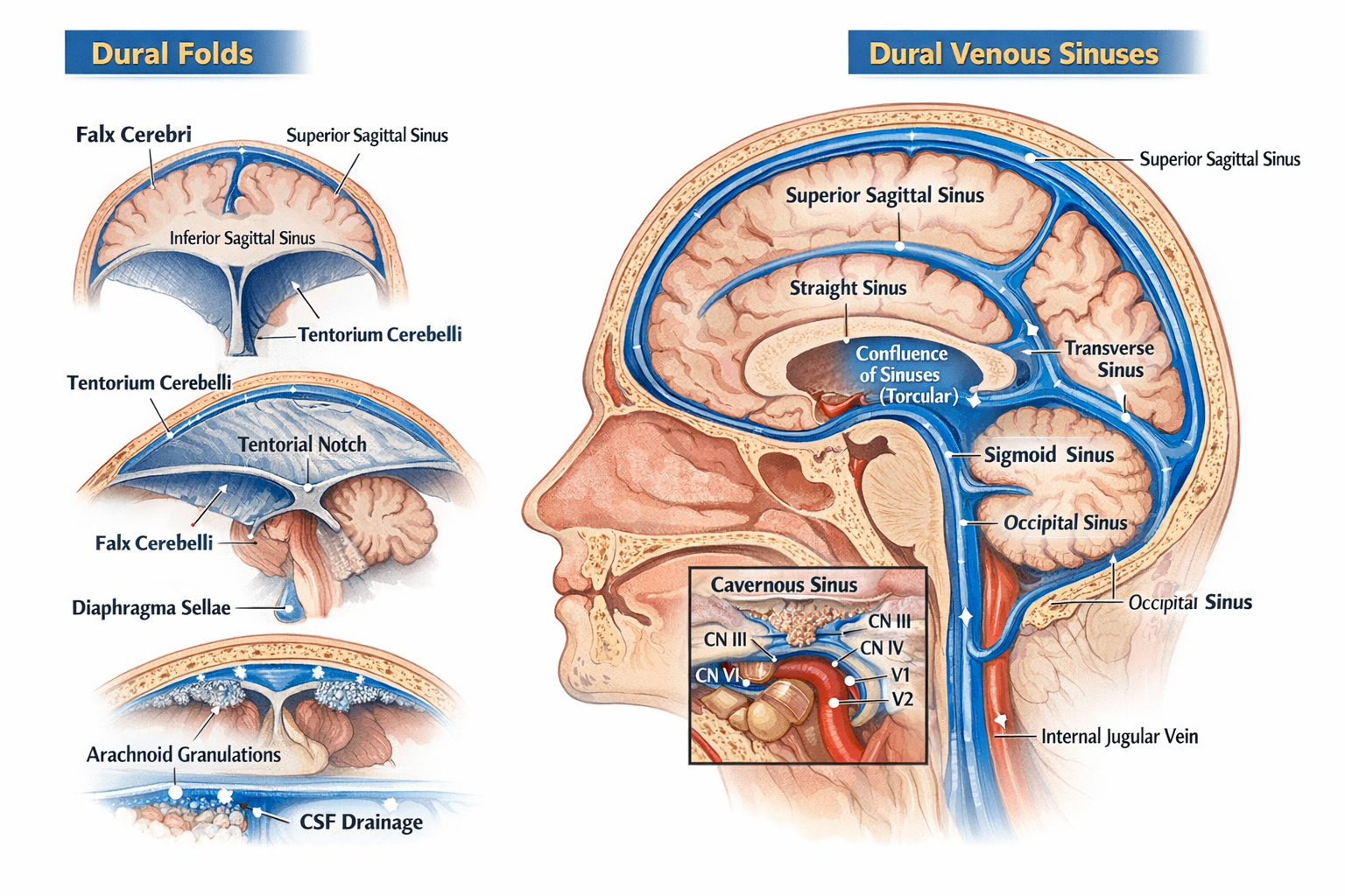
2. Dural Folds (Dural Septa)
Dural folds are double-layered reflections of meningeal dura mater that:
- Stabilize the brain
- Limit excessive movement
- Contain venous sinuses at their attachments
2.1 Falx Cerebri
A sickle-shaped vertical fold in the midline.
Attachments
- Anterior: Crista galli of ethmoid
- Posterior: Internal occipital protuberance (joins tentorium cerebelli)
Separates
- Right and left cerebral hemispheres
Venous Sinuses Contained
- Superior sagittal sinus (upper border)
- Inferior sagittal sinus (free lower border)
- Straight sinus (posterior attachment)
Clinical relevance
- Falx meningioma
- Subfalcine herniation
🔗 See also: Cerebral Herniation Syndromes
2.2 Tentorium Cerebelli
A horizontal tent-like fold.
Attachments
- Anterior: Clinoid processes
- Lateral: Superior border of petrous temporal bone
- Posterior: Occipital bone
Separates
- Cerebrum (above) from cerebellum (below)
Opening
- Tentorial notch → passage for midbrain
Venous Sinuses Contained
- Transverse sinus
- Superior petrosal sinus
- Straight sinus (junction with falx cerebri)
Clinical relevance
- Transtentorial (uncal) herniation
- Compression of oculomotor nerve
🔗 Related topic: Midbrain Anatomy
2.3 Falx Cerebelli
A small vertical fold below the tentorium.
Attachments
- Internal occipital crest
Separates
- Two cerebellar hemispheres (partially)
Venous Sinus
- Occipital sinus
2.4 Diaphragma Sellae
A small circular dural fold forming the roof of sella turcica.
Central opening
- Allows passage of pituitary stalk (infundibulum)
Clinical relevance
- Pituitary adenoma expansion
- CSF leak after trans-sphenoidal surgery
🔗 See also: Pituitary Gland Anatomy
3. Dural Venous Sinuses
Definition
Endothelial-lined venous channels between layers of dura mater that drain venous blood from brain, meninges, and skull.
Key characteristics
- No valves
- No muscular layer
- Rigid walls (do not collapse)
4. Classification of Dural Venous Sinuses
4.1 Unpaired Sinuses
Superior Sagittal Sinus
- Location: Upper margin of falx cerebri
- Drains: Cerebral veins, CSF via arachnoid granulations
- Ends in: Confluence of sinuses
Clinical
- Site of CSF absorption
- Thrombosis → raised intracranial pressure
Inferior Sagittal Sinus
- Location: Free lower margin of falx cerebri
- Drains into: Straight sinus
Straight Sinus
- Formed by union of inferior sagittal sinus + great cerebral vein (of Galen)
- Ends in: Confluence of sinuses
Occipital Sinus
- Smallest sinus
- Located in falx cerebelli
4.2 Paired Sinuses
Transverse Sinuses
- Located along posterolateral margin of tentorium
- Drain into: Sigmoid sinuses
Sigmoid Sinuses
- S-shaped
- Continue as: Internal jugular veins
🔗 Related topic: Internal Jugular Vein
Cavernous Sinus (Highly Important)
Located on either side of body of sphenoid.
Contents
- Internal carotid artery
- CN VI (abducent nerve)
Lateral wall (superior to inferior)
- CN III
- CN IV
- V1
- V2
Drains
- Superior & inferior ophthalmic veins
Clinical
- Cavernous sinus thrombosis
- Carotid–cavernous fistula
🔗 See also: Cranial Nerves in Cavernous Sinus
Petrosal Sinuses
- Superior petrosal sinus: cavernous → transverse
- Inferior petrosal sinus: cavernous → internal jugular vein
5. Confluence of Sinuses (Torcular Herophili)
- Located at internal occipital protuberance
- Receives:
* Superior sagittal sinus
* Straight sinus
* Occipital sinus
- Drains into: Transverse sinuses
6. CSF Drainage and Arachnoid Granulations
- CSF absorbed into superior sagittal sinus
- Via arachnoid villi and granulations
🔗 Related topic: CSF Circulation and Absorption
7. Clinical Correlation Summary
| Condition | Related Structure |
| ----------------- | -------------------------- |
| Raised ICP | Superior sagittal sinus |
| Uncal herniation | Tentorium cerebelli |
| Diplopia | Cavernous sinus (CN VI) |
| CSF leak | Diaphragma sellae |
| Venous thrombosis | Sagittal / cavernous sinus |
8. One-Line Exam Pearls
- Dural folds are formed by meningeal dura only
- Venous sinuses lack valves and smooth muscle
- Cavernous sinus is the only sinus containing cranial nerves
- Falx cerebri contains two sagittal sinuses