Deep Cervical Fascia and Triangles of the Neck — Detailed Anatomical Guide
Deep Cervical Fascia
Definition
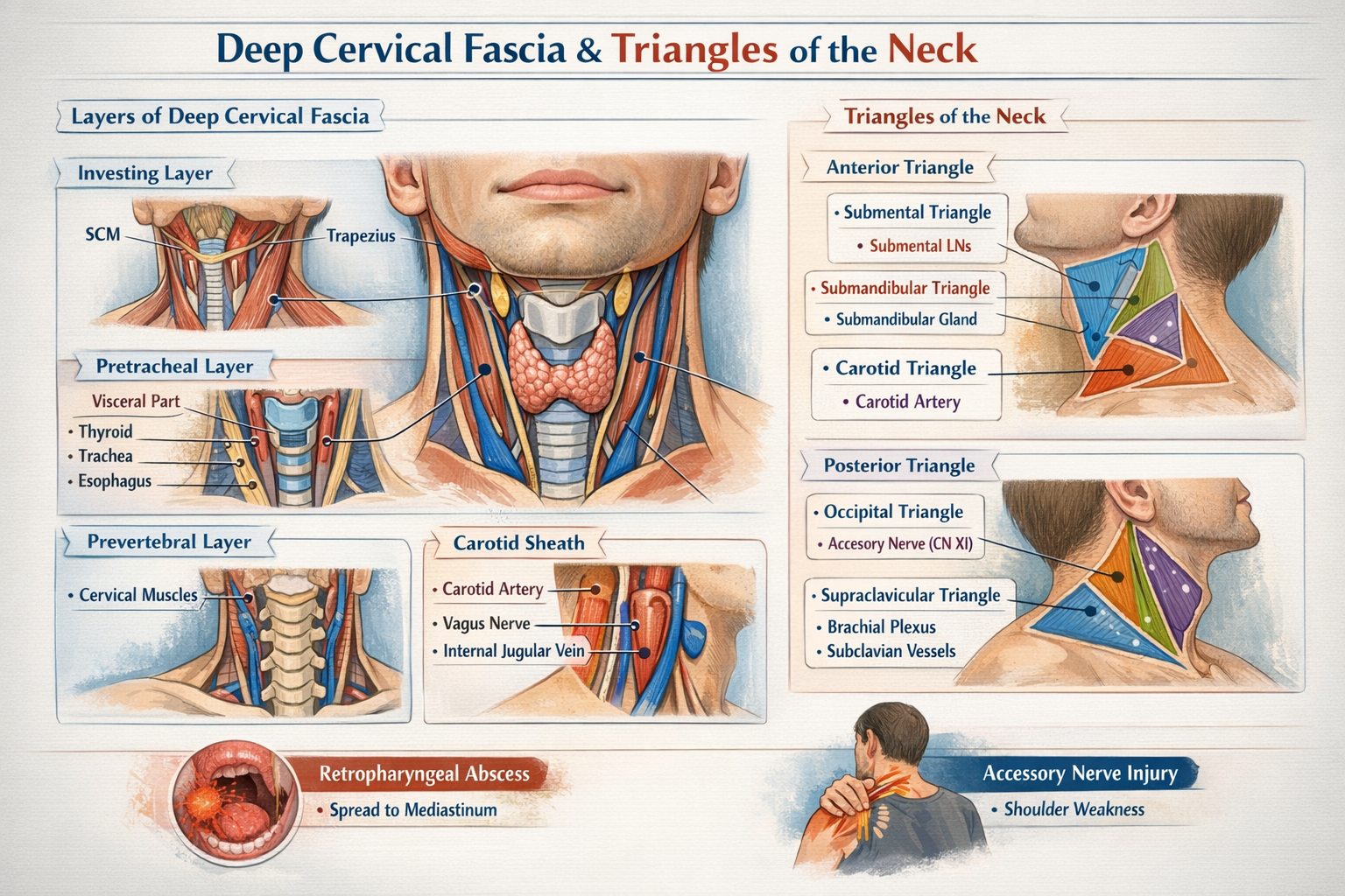
The deep cervical fascia is a dense connective tissue layer in the neck that surrounds, supports, and compartmentalizes muscles, vessels, nerves, and viscera. It plays a critical role in structural support, movement coordination, and containment of infections.
Layers of Deep Cervical Fascia
1. Investing Layer (Superficial Layer of Deep Fascia)
Extent
- Encloses the entire neck like a collar
- Splits to surround sternocleidomastoid (SCM) and trapezius
- Extends:
* Superiorly: superior nuchal line, mandible, zygomatic arch
* Inferiorly: clavicle, sternum, acromion
Attachments
- Mastoid process
- External occipital protuberance
- Lower border of mandible
- Spine of scapula
Structures Enclosed
- SCM
- Trapezius
- Parotid gland (forms parotid fascia)
- Submandibular gland (forms submandibular fascia)
Clinical Importance
- Limits superficial spread of infection
- Parotid abscess causes severe pain due to tight fascia
- Forms stylomandibular ligament
2. Pretracheal Layer
Divided into muscular and visceral parts.
A. Muscular Part
Encloses
- Infrahyoid (strap) muscles:
* Sternohyoid
* Sternothyroid
* Thyrohyoid
* Omohyoid
Extent
- Hyoid bone → superior mediastinum
B. Visceral Part
Encloses
- Thyroid gland
- Trachea
- Esophagus
Special Features
- Forms false capsule of thyroid
- Thickened posteriorly to form Berry’s ligament (anchors thyroid to cricoid cartilage)
Clinical Importance
- Explains movement of thyroid gland during swallowing
- Thyroid swelling moves with deglutition
3. Prevertebral Layer
Extent
- Base of skull → T3 vertebra
Encloses
- Cervical vertebrae
- Deep neck muscles:
* Longus colli
* Longus capitis
* Scalene muscles
- Vertebral vessels
- Cervical sympathetic trunk
Lateral Extension
- Forms axillary sheath, enclosing:
* Subclavian artery
* Brachial plexus
Clinical Importance
- Infection here can spread to posterior mediastinum
- Involvement affects neck movements
4. Carotid Sheath
A tubular condensation of deep cervical fascia formed by:
- Investing layer
- Pretracheal layer
- Prevertebral layer
Extent
- Base of skull → root of neck
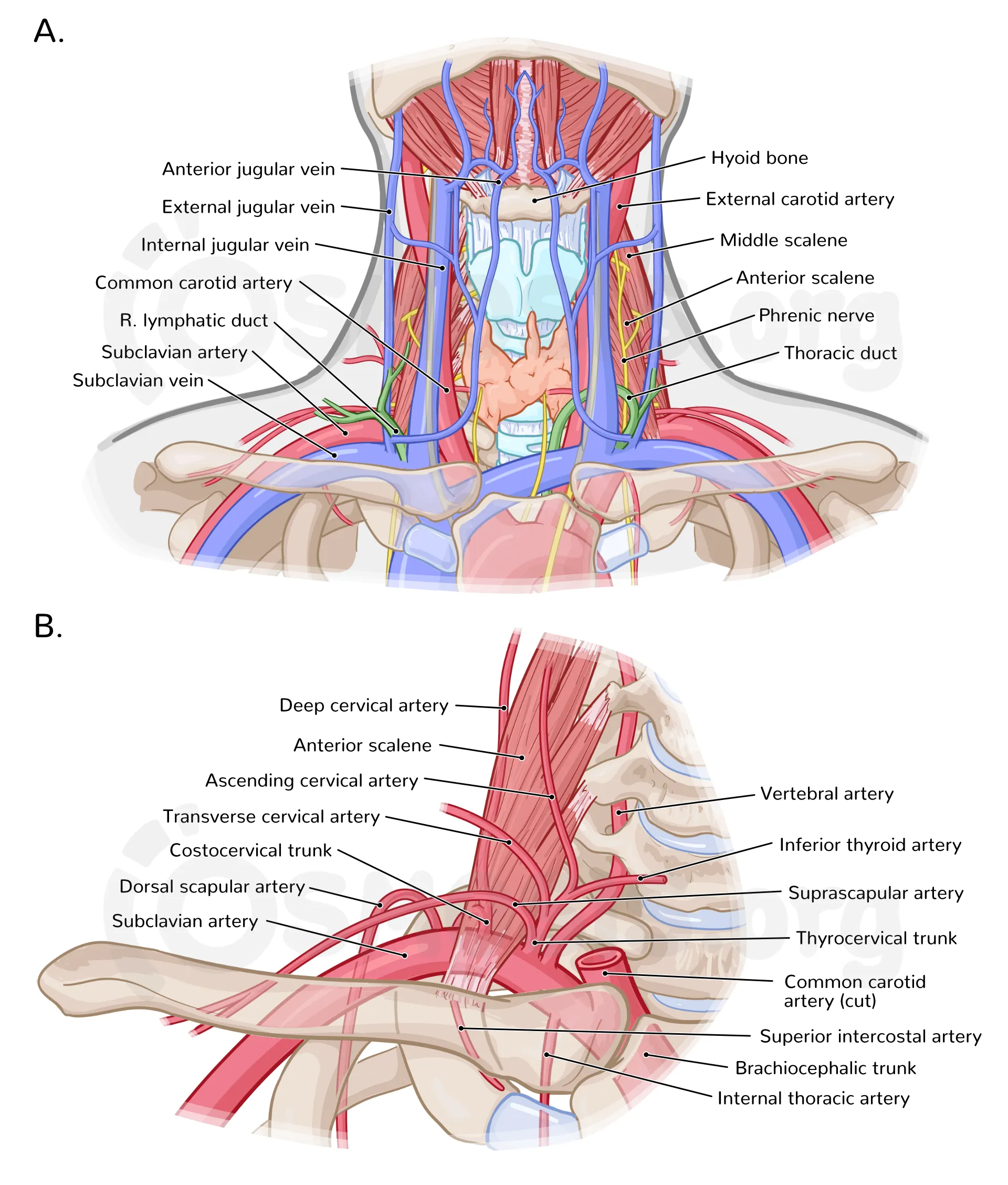
Contents
- Common carotid artery (internal carotid above bifurcation)
- Internal jugular vein
- Vagus nerve
- Deep cervical lymph nodes
- Sympathetic fibers
Arrangement
- Artery: medial
- Vein: lateral
- Nerve: posterior
Clinical Importance
- Compression can affect cerebral blood flow
- Infections can spread vertically
Spaces Formed by Deep Cervical Fascia
- Pretracheal space → anterior mediastinum
- Retropharyngeal space → posterior mediastinum (danger space)
- Prevertebral space → posterior mediastinum
Triangles of the Neck
The neck is divided by sternocleidomastoid (SCM) into anterior and posterior triangles.
Anterior Triangle
Boundaries
- Medial: midline of neck
- Lateral: anterior border of SCM
- Superior: lower border of mandible
- Apex: suprasternal notch
Roof
- Skin
- Superficial fascia
- Platysma
- Investing layer of deep fascia
Floor
- Pharynx
- Larynx
- Thyroid gland
Subdivisions of Anterior Triangle
1. Submental Triangle
Boundaries
- Two anterior bellies of digastric
- Base: body of hyoid
Contents
- Submental lymph nodes
- Small veins forming anterior jugular vein
Clinical Importance
- Drains lower lip, chin, tip of tongue
2. Submandibular (Digastric) Triangle
Boundaries
- Anterior and posterior bellies of digastric
- Lower border of mandible
Contents
- Submandibular gland
- Facial artery and vein
- Hypoglossal nerve
- Submandibular lymph nodes
3. Carotid Triangle
Boundaries
- Posterior belly of digastric
- Superior belly of omohyoid
- Anterior border of SCM
Contents
- Common carotid artery and bifurcation
- Internal and external carotid arteries
- Internal jugular vein
- Vagus nerve
- Hypoglossal nerve
- Carotid sinus and body
Clinical Importance
- Site for carotid pulse
- Carotid endarterectomy
4. Muscular Triangle
Boundaries
- Midline of neck
- Anterior border of SCM
- Superior belly of omohyoid
Contents
- Infrahyoid muscles
- Thyroid and parathyroid glands
- Larynx
- Trachea
Posterior Triangle
Boundaries
- Anterior: posterior border of SCM
- Posterior: anterior border of trapezius
- Inferior: clavicle
- Apex: where SCM and trapezius meet
Roof
- Skin
- Superficial fascia
- Platysma
- Investing layer of deep fascia
Floor
- Splenius capitis
- Levator scapulae
- Scalene muscles
Subdivision of Posterior Triangle
1. Occipital Triangle
Boundaries
- SCM
- Trapezius
- Inferior belly of omohyoid
Contents
- Spinal accessory nerve (CN XI)
- Cervical plexus branches
- Occipital artery
- Lymph nodes
Clinical Importance
- CN XI injury causes shoulder droop
2. Supraclavicular (Subclavian) Triangle
Boundaries
- Clavicle
- SCM
- Inferior belly of omohyoid
Contents
- Subclavian artery and vein
- Brachial plexus trunks
- Supraclavicular lymph nodes
Clinical Importance
- Venous access
- Pancoast tumor involvement
Key Clinical Correlations (High-Yield)
- Deep cervical fascia directs spread of neck infections
- Retropharyngeal abscess can descend into mediastinum
- Thyroid movement with swallowing explained by pretracheal fascia
- Accessory nerve vulnerability in posterior triangle
- Carotid sheath protects vital neurovascular structures