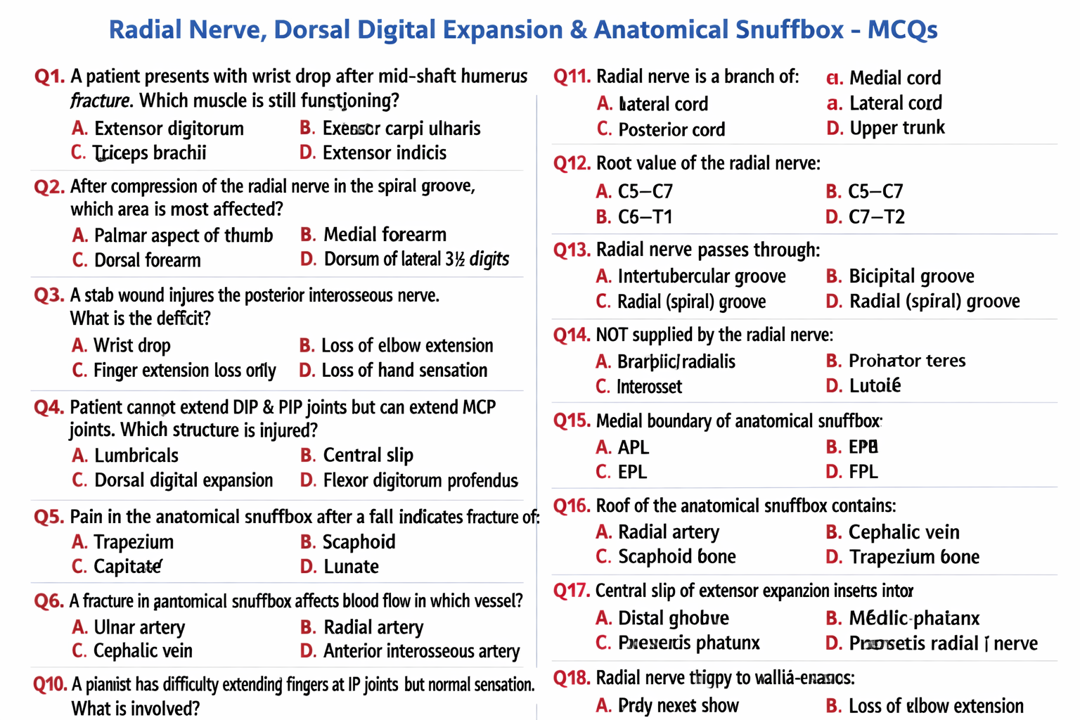
Flexor Retinaculum (Transverse Carpal Ligament) — Detailed Anatomy
Definition
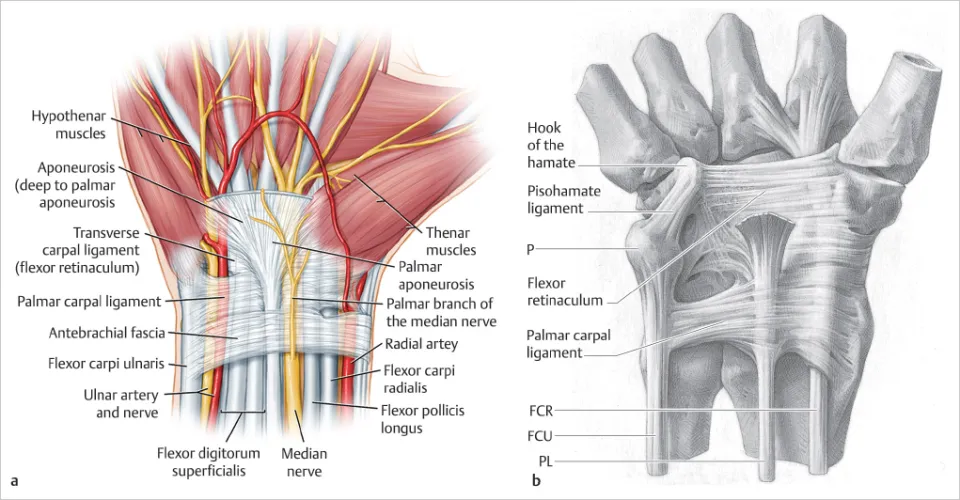
The flexor retinaculum is a strong fibrous band on the palmar aspect of the wrist. It spans the concavity of the carpal bones and converts the carpal groove into the carpal tunnel, retaining the long flexor tendons and neurovascular structures in position.
Location & Extent
- Situated across the anterior (palmar) wrist
- Forms the roof of the carpal tunnel
- Lies superficial to the carpal bones and deep to palmar skin and fascia
Attachments
Medial (ulnar) side
- Pisiform
- Hook of hamate
Lateral (radial) side
- Tubercle of scaphoid
- Crest/tubercle of trapezium
Structure & Relations
- Thickened part of deep fascia of the forearm
- Continuous proximally with deep fascia
- Gives attachment to thenar and hypothenar muscles
- Superficial to flexor tendons and median nerve
- Palmar aponeurosis lies superficial to it
Contents Beneath the Flexor Retinaculum (Carpal Tunnel Contents)
Within the carpal tunnel (deep to retinaculum):
- Median nerve
- 9 flexor tendons:
* 4 tendons of flexor digitorum superficialis
* 4 tendons of flexor digitorum profundus
* 1 tendon of flexor pollicis longus
Outside the tunnel but related:
- Flexor carpi radialis tendon (in its own compartment)
- Ulnar nerve and ulnar artery (pass superficial to retinaculum through Guyon’s canal)
- Palmar cutaneous branch of median nerve (superficial)
Functions
- Prevents bowstringing of flexor tendons during wrist flexion
- Maintains mechanical efficiency of finger flexors
- Forms a protective fibro-osseous tunnel for median nerve and tendons
Clinical Importance
Carpal Tunnel Syndrome
- Compression of median nerve beneath flexor retinaculum
- Symptoms: pain, numbness, tingling in thumb, index, middle, and lateral half of ring finger; thenar muscle weakness
Surgical Relevance
- Flexor retinaculum release relieves median nerve compression
- Care taken to avoid injury to:
* Recurrent branch of median nerve
* Superficial palmar arch
Applied Anatomy Points (Exam-Oriented)
- Median nerve lies deep to retinaculum, palmar cutaneous branch is superficial
- Ulnar nerve is not compressed in carpal tunnel syndrome
- Thickening or edema of retinaculum worsens tunnel pressure