Blood Supply of the Brain
Introduction
The brain, despite forming only about 2% of total body weight, receives nearly 20–25% of cardiac output. Continuous cerebral perfusion is essential because neurons have high metabolic demand and minimal energy storage. The blood supply of the brain is designed with dual arterial systems and collateral circulation to protect against ischemia.
Arterial Supply of the Brain
The arterial supply is divided into two major systems:
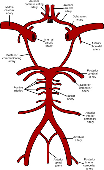
1. Internal Carotid Arterial System (Anterior Circulation)
Supplies approximately two-thirds of the cerebral hemispheres.
Origin
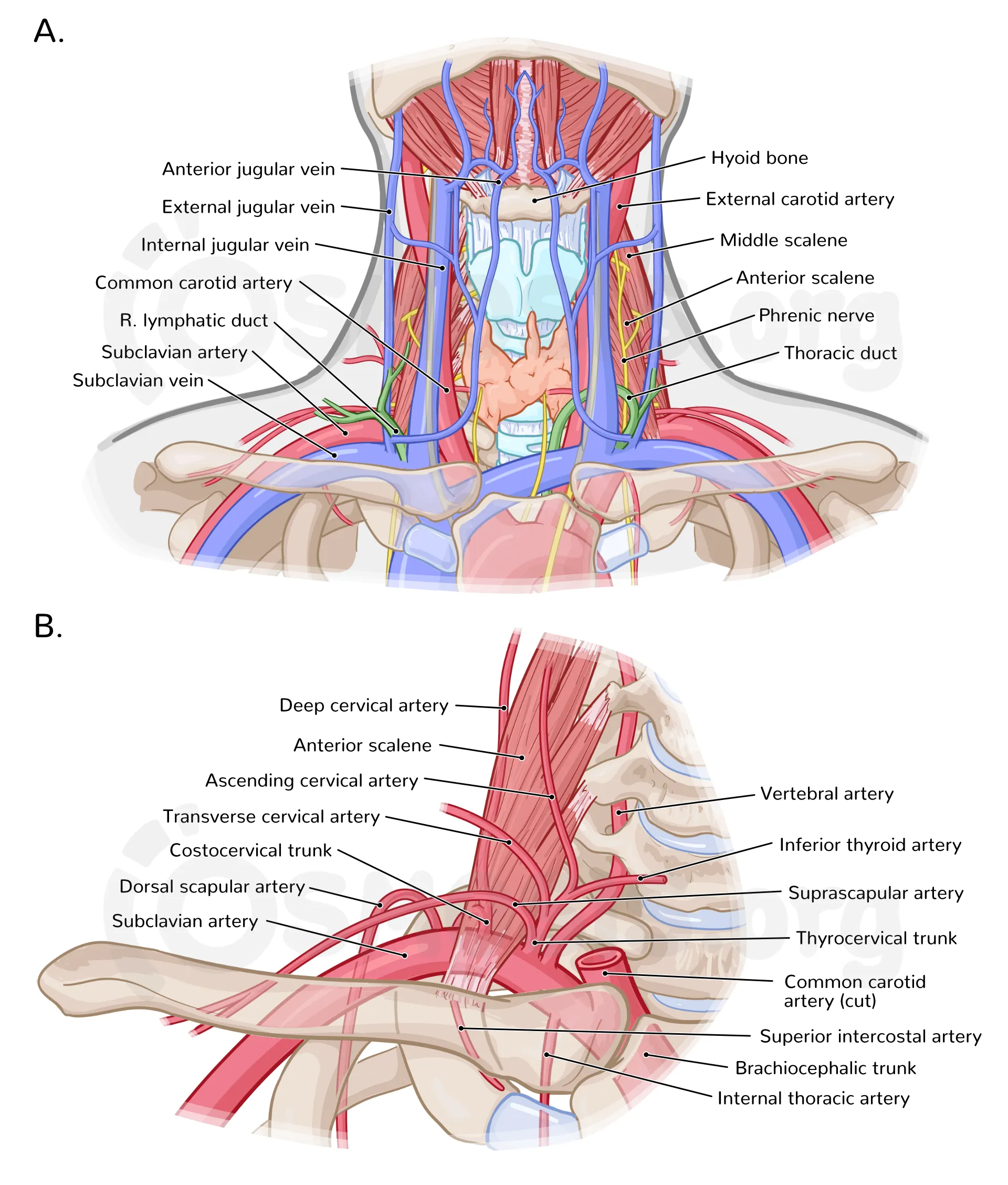
- Common carotid artery → Internal carotid artery (ICA)
Major Branches of ICA
- Anterior cerebral artery (ACA)
- Middle cerebral artery (MCA)
- Posterior communicating artery
- Anterior choroidal artery
Areas Supplied
- Frontal lobe (motor, behavior)
- Parietal lobe (sensory)
- Lateral temporal lobe (language)
- Basal ganglia and internal capsule (via lenticulostriate branches)
2. Vertebrobasilar System (Posterior Circulation)
Supplies the brainstem, cerebellum, occipital lobes, and part of temporal lobes.
Origin
- Subclavian artery → Vertebral arteries → Basilar artery
Branches
- Posterior inferior cerebellar artery (PICA)
- Anterior inferior cerebellar artery (AICA)
- Superior cerebellar artery (SCA)
- Posterior cerebral artery (PCA)
Areas Supplied
- Brainstem (midbrain, pons, medulla)
- Cerebellum
- Occipital lobe (visual cortex)
- Inferior temporal lobe
Circle of Willis
A hexagonal arterial anastomotic ring located at the base of the brain.
Components
- Anterior cerebral arteries (right and left)
- Anterior communicating artery
- Internal carotid arteries
- Posterior communicating arteries
- Posterior cerebral arteries
Functions
- Provides collateral circulation
- Maintains cerebral perfusion during arterial blockage
- Equalizes pressure between anterior and posterior circulation
Cortical Arterial Territories
Anterior Cerebral Artery (ACA)
- Medial frontal and parietal lobes
- Motor and sensory areas for contralateral lower limb
Middle Cerebral Artery (MCA)
- Lateral surface of cerebral hemispheres
- Motor and sensory cortex for face and upper limb
- Dominant hemisphere → speech areas
Posterior Cerebral Artery (PCA)
- Occipital lobe
- Visual cortex
- Inferior temporal lobe
Deep Cerebral Blood Supply
Supplied by perforating arteries:
- Lenticulostriate arteries (from MCA)
- Thalamoperforators (from PCA)
Structures Supplied
- Basal ganglia
- Internal capsule
- Thalamus
These vessels are end arteries, highly prone to lacunar infarcts.
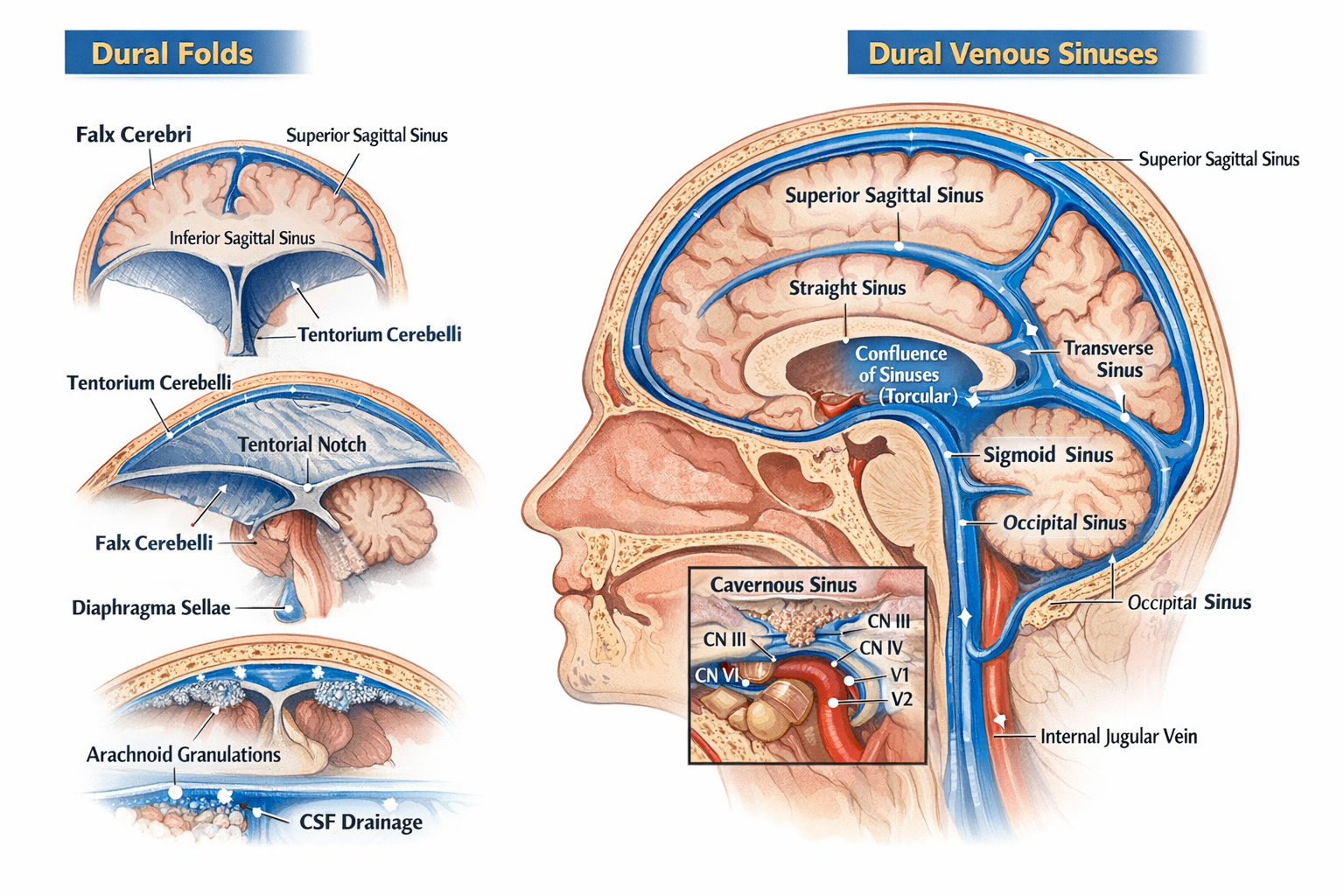
Venous Drainage of the Brain
Superficial Cerebral Veins
- Drain cerebral cortex
- Empty into dural venous sinuses
Deep Cerebral Veins
- Internal cerebral veins
- Great cerebral vein (vein of Galen)
Dural Venous Sinuses
- Superior sagittal sinus
- Inferior sagittal sinus
- Straight sinus
- Transverse sinus
- Sigmoid sinus → Internal jugular vein
Regulation of Cerebral Blood Flow
- Autoregulation maintains constant flow (MAP 60–160 mmHg)
- Increased CO₂ → Vasodilation
- Decreased O₂ → Vasodilation
- Sympathetic tone has minimal effect
Clinical Correlation
Common Stroke Territories
- MCA stroke: Contralateral hemiplegia (face > leg), aphasia
- ACA stroke: Contralateral leg weakness, behavioral changes
- PCA stroke: Visual field defects
Other Conditions
- Berry aneurysm (Circle of Willis)
- Hypertensive hemorrhage (lenticulostriate arteries)
- Vertebrobasilar insufficiency
Key Points for Exams
- Brain receives 750 mL/min blood flow
- MCA is most commonly occluded artery
- Lenticulostriate arteries are end arteries
- Circle of Willis provides collateral circulation