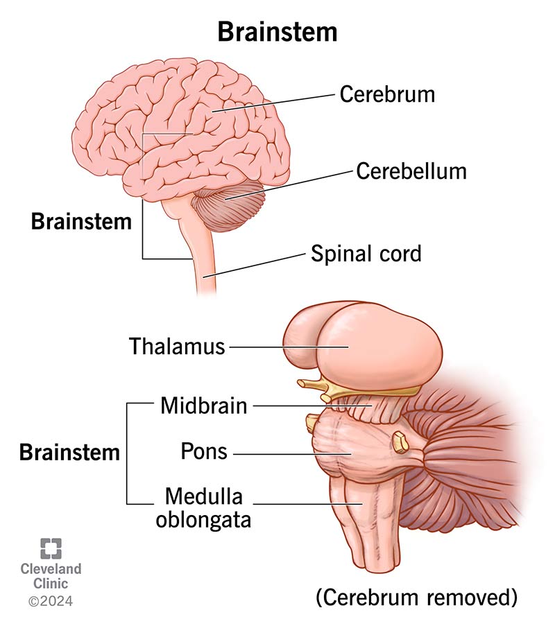
Components of the Brainstem
The brainstem consists of three parts (from above downward):
- Midbrain (Mesencephalon)
- Pons
- Medulla Oblongata
1. Midbrain (Mesencephalon)
Location
- Between the pons and diencephalon
- Traversed by the cerebral aqueduct
External Features
- Ventral: Cerebral peduncles
- Dorsal: Superior and inferior colliculi (tectum)
Internal Structure
- Tectum – Visual and auditory reflex centers
- Tegmentum – Red nucleus, periaqueductal gray
- Basis pedunculi – Motor tracts
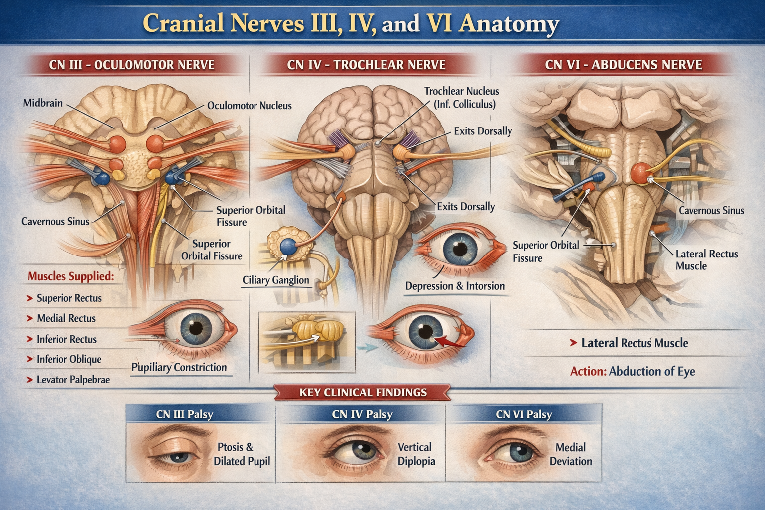
Cranial Nerve Nuclei
- CN III (Oculomotor)
- CN IV (Trochlear)
Functions
- Eye movements
- Visual and auditory reflexes
- Motor control modulation
2. Pons
Location
- Between midbrain and medulla
- Anterior to cerebellum
External Features
- Basilar groove for basilar artery
- Middle cerebellar peduncles
Internal Structure
- Basal part: Pontine nuclei, transverse fibers
- Tegmental part: Cranial nerve nuclei, tracts
Cranial Nerve Nuclei
- CN V (Trigeminal)
- CN VI (Abducens)
- CN VII (Facial)
- CN VIII (Vestibulocochlear – part)
Functions
- Relay between cerebrum and cerebellum
- Regulation of respiration
- Facial sensation and expression
3. Medulla Oblongata
Location
- From pons to spinal cord
- Ends at foramen magnum
External Features
- Pyramids – Motor tracts
- Olives – Inferior olivary nucleus
Internal Structure
- Open (upper) and closed (lower) medulla
- Nucleus gracilis and cuneatus
Cranial Nerve Nuclei
- CN VIII (part)
- CN IX (Glossopharyngeal)
- CN X (Vagus)
- CN XI (Accessory)
- CN XII (Hypoglossal)
Functions
- Cardiac and respiratory control
- Swallowing, coughing, vomiting reflexes
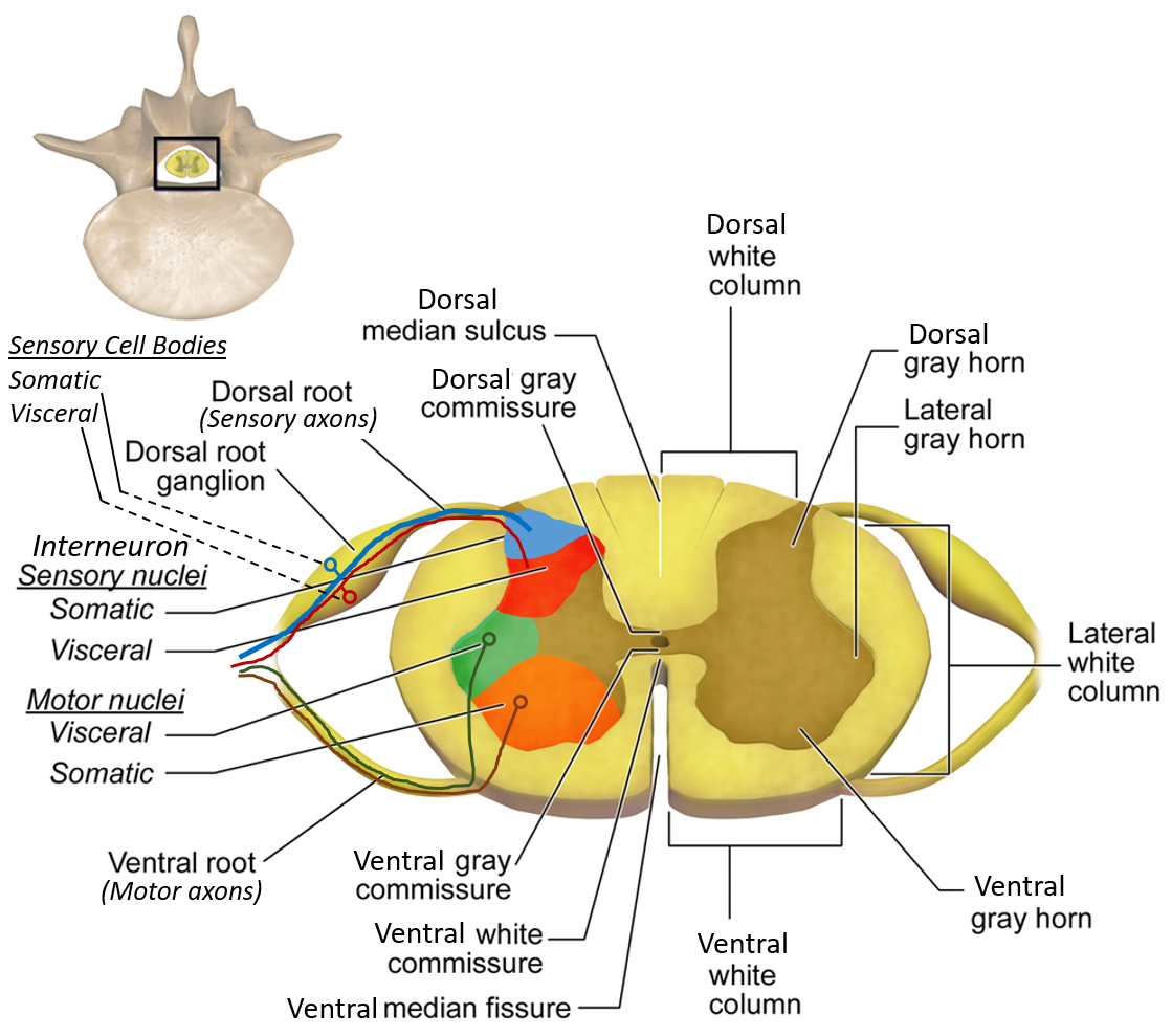
Cranial Nerve Nuclei Organization
Brainstem nuclei are arranged longitudinally:
- Medial: Motor nuclei
- Lateral: Sensory nuclei
Functional Columns
- GSE – Somatic motor
- GVE – Autonomic motor
- GSA – Somatic sensory
- GVA – Visceral sensory
Ascending (Sensory) Tracts
- Medial lemniscus – Fine touch, proprioception
- Spinothalamic tract – Pain and temperature
- Spinocerebellar tracts – Unconscious proprioception
Descending (Motor) Tracts
- Corticospinal tract – Voluntary motor control
- Corticobulbar tract – Cranial nerve motor control
- Rubrospinal, vestibulospinal tracts – Posture and tone
Reticular Formation
- Diffuse neuronal network
- Regulates consciousness, sleep-wake cycle, muscle tone, autonomic control
Blood Supply of Brainstem
- Vertebral artery
- Basilar artery
- Posterior cerebral artery
- Anterior spinal artery
Clinical Correlations
- Midbrain lesions: Weber, Benedikt syndromes
- Pontine lesions: Facial paralysis, locked-in syndrome
- Medullary lesions: Lateral medullary (Wallenberg) syndrome
- Brainstem death: Loss of vital reflexes and consciousness
Importance in Clinical Practice
- Damage can be life-threatening
- Explains cranial nerve palsies and crossed neurological signs
- Critical in stroke localization and neuroimaging interpretation
Frequently Asked Questions (FAQ)
What is the main function of the brainstem?
It controls vital functions such as breathing, heart rate, consciousness, and cranial nerve activity.
Which cranial nerves arise from the brainstem?
All cranial nerves except CN I (Olfactory) and CN II (Optic).
Why are brainstem lesions dangerous?
Because the brainstem contains respiratory and cardiovascular centers essential for survival.
Which part of the brainstem controls respiration?
Primarily the pons and medulla oblongata.
What causes crossed paralysis in brainstem lesions?
Motor fibers cross at different levels, producing ipsilateral cranial nerve deficits and contralateral body weakness.