Medical Disclaimer: This is educational content only, not medical advice. Consult a licensed healthcare provider for diagnosis/treatment. Information based on sources like WHO/CDC guidelines (last reviewed: 2026-02-13).
This article is being expanded for more depth. Check back soon!
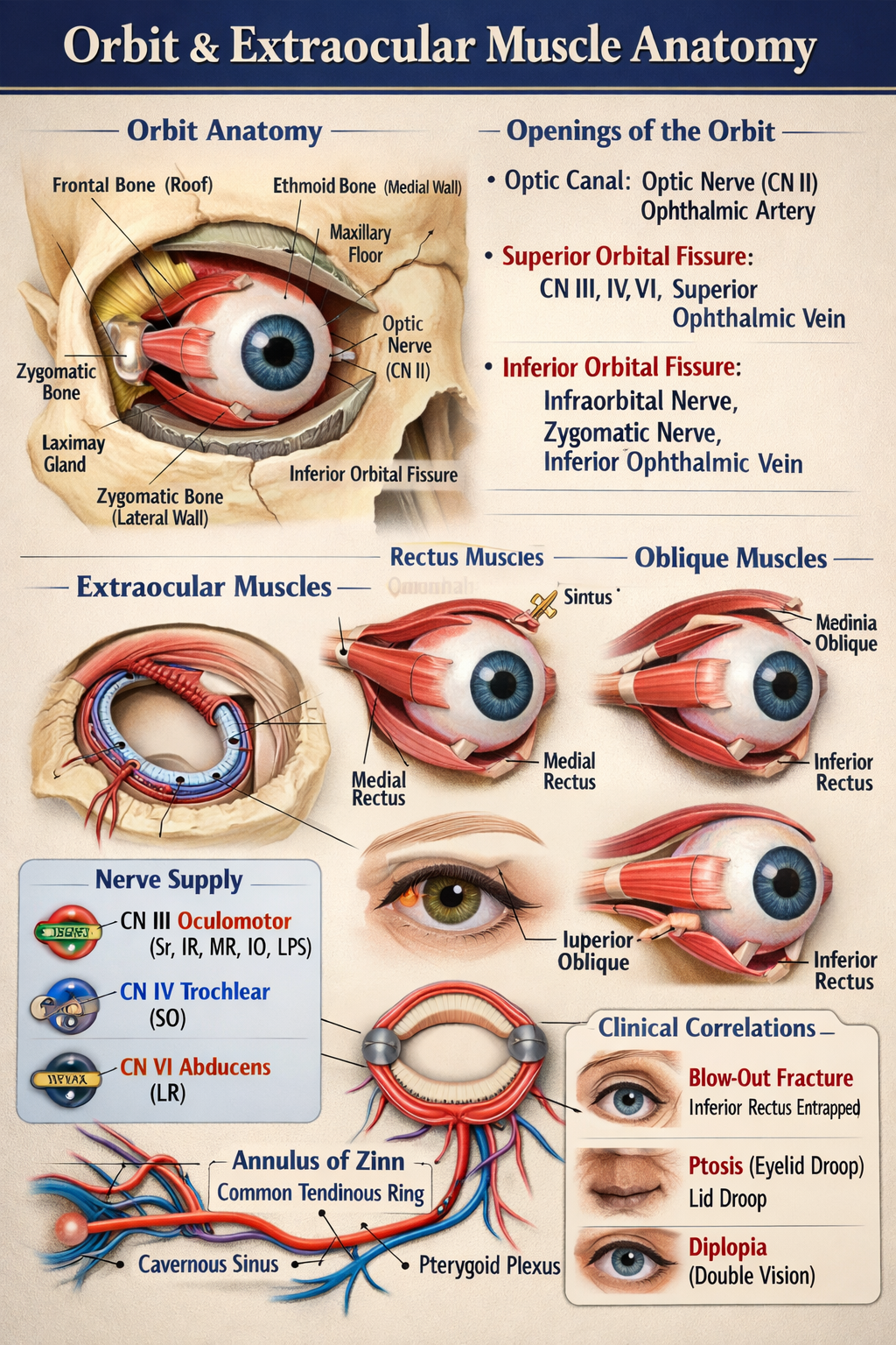
Orbit and Extraocular Muscle Anatomy Explained for Medical Students
Frequently Asked Questions
What is the orbit in human anatomy?
The orbit is a pyramidal bony cavity of the skull that contains the eyeball, extraocular muscles, optic nerve, blood vessels, lacrimal gland, and supporting connective tissue.
How many bones form the bony orbit?
The bony orbit is formed by seven bones: frontal, sphenoid, zygomatic, maxilla, palatine, ethmoid, and lacrimal bones.
What forms the roof of the orbit?
The roof of the orbit is formed by the frontal bone and the lesser wing of the sphenoid, separating the orbit from the anterior cranial fossa.
Which wall of the orbit is the weakest and why?
The medial wall is the weakest because it is formed mainly by the lamina papyracea of the ethmoid bone, making it thin and prone to infection spread from the ethmoidal sinuses.
What are the main openings of the orbit?
The main openings are the optic canal, superior orbital fissure, and inferior orbital fissure, which transmit nerves and vessels between the orbit and cranial fossae.
What structures pass through the optic canal?
The optic canal transmits the optic nerve (cranial nerve II) and the ophthalmic artery.
How many extraocular muscles are present and what are they?
There are seven extraocular muscles: four recti (superior, inferior, medial, lateral), two obliques (superior and inferior), and one levator palpebrae superioris.
What is the annulus of Zinn?
The annulus of Zinn is a common tendinous ring located at the orbital apex from which all four rectus muscles and the levator palpebrae superioris originate.
Which nerve supplies most of the extraocular muscles?
The oculomotor nerve (cranial nerve III) supplies all extraocular muscles except the superior oblique and lateral rectus.
What is a blow-out fracture of the orbit?
A blow-out fracture is a fracture of the orbital floor, usually involving the maxilla, which can trap the inferior rectus muscle and cause diplopia.
MCQ Test - Orbit and Extraocular Muscle Anatomy Explained for Medical Students
Progress:
0/0
Time: 00:00
No MCQs available for this article.