Medical Disclaimer: This is educational content only, not medical advice. Consult a licensed healthcare provider for diagnosis/treatment. Information based on sources like WHO/CDC guidelines (last reviewed: 2026-02-13).
This article is being expanded for more depth. Check back soon!
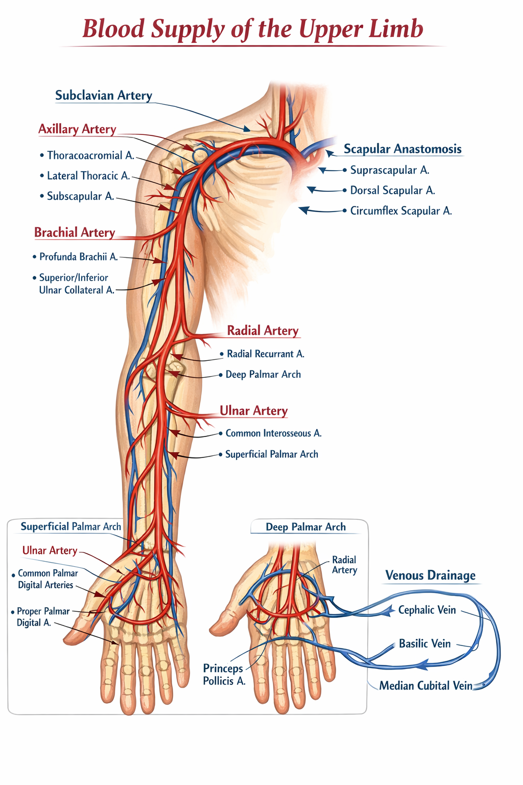
Blood Supply of Upper Limb Anatomy Explained With Clinical Importance
Frequently Asked Questions
What is the main arterial source of blood supply to the upper limb?
The main arterial source of blood supply to the upper limb is the subclavian artery, which continues as the axillary artery and then the brachial artery.
At what point does the subclavian artery become the axillary artery?
The subclavian artery becomes the axillary artery at the lateral border of the first rib.
How is the axillary artery divided anatomically?
The axillary artery is divided into three parts by the pectoralis minor muscle: first part proximal, second part posterior, and third part distal to the muscle.
Which is the largest branch of the axillary artery?
The subscapular artery is the largest branch of the axillary artery.
Which artery accompanies the radial nerve in the spiral groove of the humerus?
The profunda brachii artery accompanies the radial nerve in the spiral groove of the humerus.
Where does the brachial artery terminate?
The brachial artery terminates in the cubital fossa by dividing into the radial and ulnar arteries.
Which artery is commonly used for measuring pulse at the wrist?
The radial artery is commonly used for measuring pulse at the wrist.
Which artery mainly forms the superficial palmar arch?
The superficial palmar arch is mainly formed by the ulnar artery.
Which artery mainly forms the deep palmar arch?
The deep palmar arch is mainly formed by the radial artery.
What is the main function of scapular anastomosis?
Scapular anastomosis provides collateral circulation to the upper limb in cases of axillary artery blockage.
Which vein is commonly used for venepuncture in the upper limb?
The median cubital vein is the most commonly used vein for venepuncture.
What are venae comitantes?
Venae comitantes are paired deep veins that accompany arteries in the upper limb.
Which artery supplies the thumb primarily?
The princeps pollicis artery primarily supplies the thumb.
Which artery passes through the anatomical snuffbox?
The radial artery passes through the anatomical snuffbox.
Why is the blood supply of the upper limb clinically important?
It is clinically important for procedures such as arterial cannulation, venepuncture, fracture management, vascular surgeries, and assessment of ischemic conditions.
MCQ Test - Blood Supply of Upper Limb Anatomy Explained With Clinical Importance
Progress:
0/0
Time: 00:00
No MCQs available for this article.