Medical Disclaimer: This is educational content only, not medical advice. Consult a licensed healthcare provider for diagnosis/treatment. Information based on sources like WHO/CDC guidelines (last reviewed: 2026-02-13).
This article is being expanded for more depth. Check back soon!
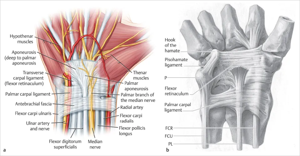
Flexor Retinaculum Anatomy Structure Attachments Functions and Clinical Importance
Frequently Asked Questions
What is the flexor retinaculum?
The flexor retinaculum is a strong fibrous band on the palmar aspect of the wrist that converts the carpal groove into the carpal tunnel and holds flexor tendons and the median nerve in place.
Where is the flexor retinaculum located?
It is located on the anterior (palmar) surface of the wrist, stretching between the carpal bones on the radial and ulnar sides.
What are the attachments of the flexor retinaculum?
Laterally it attaches to the tubercle of scaphoid and crest of trapezium, and medially to the pisiform and hook of hamate.
Which structures pass deep to the flexor retinaculum?
The median nerve and nine flexor tendons (four flexor digitorum superficialis, four flexor digitorum profundus, and one flexor pollicis longus) pass deep to it.
Which structures pass superficial to the flexor retinaculum?
The ulnar nerve, ulnar artery, and palmar cutaneous branch of the median nerve pass superficial to the flexor retinaculum.
What is the main function of the flexor retinaculum?
Its main function is to prevent bowstringing of flexor tendons during wrist and finger flexion and to maintain mechanical efficiency.
How is the flexor retinaculum related to carpal tunnel syndrome?
Thickening or increased pressure beneath the flexor retinaculum compresses the median nerve, leading to carpal tunnel syndrome.
Why is palmar skin sensation spared in carpal tunnel syndrome?
Because the palmar cutaneous branch of the median nerve passes superficial to the flexor retinaculum and is not compressed.
Does the ulnar nerve pass through the carpal tunnel?
No, the ulnar nerve passes superficial to the flexor retinaculum through Guyon’s canal.
What happens during surgical release of the flexor retinaculum?
The retinaculum is cut to increase the volume of the carpal tunnel and relieve pressure on the median nerve.
MCQ Test - Flexor Retinaculum Anatomy Structure Attachments Functions and Clinical Importance
Progress:
0/0
Time: 00:00
No MCQs available for this article.